


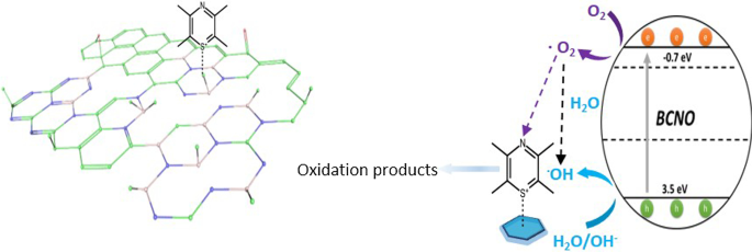
新しい金属を含まない光触媒としてのホウ素酸窒化物
要約
ホウ素ベースのナノ材料は、太陽水素燃料の製造と環境修復のための太陽エネルギー変換において、無毒で地球に豊富な(光)電極触媒材料として浮上しています。ホウ素酸窒化物(BCNO)は、電子的、光学的、および物理化学的特性を備えた4級半導体であり、ホウ素、窒素、炭素、および酸素の組成を変えることで調整できます。ただし、BCNOの構造と光触媒活性の関係はまだ調査されていません。 BCNOの調製において、2つの異なる窒素前駆体を使用した場合の影響と、アニーリング温度の影響を解明するために、詳細な分光分析を実行しました。 BCNOナノディスク( D =6.7±1.1nm)ターボストラティック窒化ホウ素回折パターンは、800°Cでの熱アニーリング時に窒素源前駆体として塩酸グアニジンを使用して調製しました。 BCNOナノディスクのX線光電子分光法(XPS)表面元素分析により、B、C、N、およびOの組成がそれぞれ40.6%、7.95%、37.7%、および13.8%であることが明らかになりました。ソリッドステートによると 11 B NMR分析では、塩酸グアニジン由来のBCNOナノディスクは、さまざまな三配位BN x の形成を示しました。 (OH) 3- x 光触媒活性部位の1つとしても機能した種。 XRDおよび詳細な分光分析により、BCNOをドープした六方晶窒化ホウ素ナノディスクの準備が裏付けられました。対照的に、窒素前駆体としてメラミンを使用して600°CでアニールされたBCNOは、XRD、XPS、および固体NMRによる証拠として、ハニカム格子に共有結合したB、C、N、およびO原子で構成される層状ナノシートで構成されていました。分析( 11 Bおよび 13 C)分析。メラミン由来のBCNO層状構造のXPS表面元素組成は、比較的低いホウ素(5.24%)と窒素(7.27%)の組成を持つ高炭素組成(75.1%)で構成され、BCNOドープ酸化グラフェンの形成を示しました。層状シート構造。この一連のメラミン由来のBCNOドープ酸化グラフェン層状構造は、グラファイト状窒化炭素の光触媒活性を超えて、最高の光触媒活性を示すことがわかりました。この層状構造では、四配位BN x の形成 (OH) 3- x (CO)種と豊富なグラファイトドメインは、BCNOをドープした酸化グラフェンの層状構造の光触媒活性に重要な役割を果たすことが提案されました。光学バンドギャップエネルギーは、BCNOをドープした六方晶窒化ホウ素ナノディスクとBCNOをドープした酸化グラフェンの層状構造でそれぞれ5.7eVと4.2eVと測定されました。最後に、BCNOは、BGH01、BGH03、BMH01、BMH03で、それぞれ1.58、2.10、5.18、8.14 µsの平均減衰寿命を持つ超長フォトルミネッセンスを示しました。この研究は、新しい金属を含まない光触媒システムを提供し、BCNOベースの光触媒の起源に関する最初の構造分析を提供します。
グラフィカルな要約
はじめに
金属を含まないナノ材料は、太陽燃料の生産、環境修復、CO 2 などのさまざまな用途向けに、構造的および化学的安定性が高く、費用対効果が高く、地球に優しい(写真)触媒として登場しています。 有害な微生物の還元、消毒、および有機化合物の選択的な化学合成を可能にしました[1,2,3,4,5,6,7]。それらの金属対応物と比較して、金属を含まない触媒はまた、中毒を起こしにくく、より長いサイクル寿命をもたらす。したがって、安定性があり、効率的で、費用効果の高い光触媒の残骸である新しい材料の探索と開発は、重要で挑戦的な研究努力です。グラファイト状窒化炭素(CN)[4、8、9]、カーボンドット(C-dot)[2、3、10]、グラフェンベースの材料[7、11]などのカーボンベースの材料が広く研究されています。それらの優れた物理化学的特性、構造的および化学的安定性、および地球に豊富な元素からの合成の容易さのために。最近、ホウ素ベースの(光)触媒が、優れた性能を備えた金属を含まない光触媒システムとして開発されました。特に、硬度で知られる炭化ホウ素は、金属を含まない可視光光触媒水素生成を示し、最先端の炭素ベースのCN光触媒を上回りました[12、13]。 H 2 に対して可視光光触媒活性を示す高表面積の炭素ドープ六方晶窒化ホウ素(BCN)ナノシート andO 2 世代とCO 2 縮小とキャプチャにより、フォトシステムに新しい可能性が生まれました[14、15]。酸窒化ホウ素(BNO)[16、17]、リン酸ホウ素(BP)[18、19]、ホウ素ドープグラフェン[20]、窒化ホウ素ホウ素(BCN)[14]、ホウ素をドープした窒化炭素(Bをドープした CN )[21]および元素ホウ素[22、23]は、重要な(写真)電極触媒活性を示しています[13]。
ホウ素酸窒化物(BCNO)は、他の材料よりも研究が少ないホウ素ベースのナノ材料です。これは、BCNの前身である、バンドギャップが約2 eVの半導体の後に開発され、酸窒化物および窒化物化合物に基づく有毒なリン光物質に取って代わりました[24、25]。グラフェンまたは六方晶窒化ホウ素(hBN)ネットワークへのB、C、O、およびN原子の置換により、調整可能なフォトルミネッセンス特性と0 eV(グラフェン)から5.9 eV(hBN)の範囲のバンドギャップを持つBCNO化合物が生成されました[26]。 。これらの望ましい半導体およびフォトルミネッセンス特性は、最近、より高い結晶化度[27]、制御された形状[28]、および原子的に薄い2D構造[29]を備えた低次元BCNOナノ構造を合成する新しい合成方法を開発する研究者を魅了しています。以前の研究では、詳細な構造特性を提供せずに、BCNOのフォトルミネッセンス特性を調整する際のアニーリング温度と時間の影響を調査しました[30、31]。この論文では、さまざまな窒素源前駆体、焼成温度(800°C対600°C)、および焼成時間(0.5時間対12時間)を使用した場合の、BCNOナノ構造の構造-光触媒活性への影響を調査しました。
>メソッド
化学薬品および機器
ホウ酸99.99%(H 3 BO 3 )、メラミン99%(C 3 H 6 N 6 )、およびヘキサメチレンテトラミン≥99%(C 6 H 12 N 4 )はAlfa Aesarから購入し、さらに精製することなく使用しました。塩酸グアニジン99.5%(CH 5 N 3 HCl)はArcosOrganicsから購入しました。 BCNOは、文献に従って低温アニーリング法[25、32]を介して合成されました。 UPS分析は、ULVAC-PHI PHI 5000 Versaprobe IIで、He I 21.22eVを5Vバイアスの光子源として使用して実行されました。 BCNOサンプルの形態は、透過型電子顕微鏡(JEOL、JEM-ARM200FTH)を介して分析されました。 XRD回折は、BrukerD2分光計を使用して取得しました。溶液中のフォトルミネッセンス発光スペクトルは、フォトルミネッセンス分光計(PerkinElmer、LS55)を使用して取得し、溶液中のBCNOの光吸収スペクトルは、UV-Vis分光計(HITACHI、U-3900)を使用して決定しました。 X線光電子分光法は、励起源としてAl Ka X線を使用して、高分解能X線光電子分光計(ULVAC-PHI、PHI Quantera II)を介して分析されました。 BCNO溶液は、XPS特性評価のためにシリコン基板上にドロップキャストされました。結合エネルギーは、284.8eVの炭素に対して較正されました。 XPSピークデコンボリューションとフィッティングは、CACSXPSソフトウェアを使用して実行されました。絶対PLQYは文献[33]に従って実行され、CCDカメラ(PIXIS 256BR、Princeton Instruments)によって検出されました。絶対PLQYの測定は、キャリブレーションされた積分球システム(Labsphere)と電荷結合デバイス(CCD)カメラ(PIXIS 256BR、Princeton Instruments)を含むファイバーベースの分光器を使用して実行されました。ダイオードレーザー(λ =375 nm、Becker&Hickl GmbH)をポンプ源として使用しました。時間分解フォトルミネッセンスは、パルス窒素レーザー(λ)を使用して前面構成で測定されました。 =337.1 nm、LTB Lasertechnik Berlin GmbH)を励起源として使用し、デジタル遅延ジェネレーター(DG645、Stanford Research Systems)によってトリガーされました。信号は光電子増倍管(PMC-100–1、Becker&Hickl GmbH)で検出され、光子数はマルチスケーラーモジュール(MSA-300、Becker&Hickl GmbH)で累積されました。赤外線スペクトルは、減衰全反射(ATR)を使用して、フーリエ変換赤外分光計(Bruker、Vertex 80v)によって記録されました。核磁気共鳴スペクトルは、4mmのマジック角回転(MAS)プローブを使用して9.4Tの磁石を備えたBrukerAvance III 400NMR分光計を使用して取得しました。 11 B MAS NMRは、10kHzのスピン速度でスピンエコー法を使用して記録されました。 8000回のスキャンが4秒のリサイクル遅延で収集されました。化学シフトは1M H 3 を基準としました BO 3 19.6ppmの水溶液。 13 C CP / MAS NMRスペクトルは、12.5 kHzの回転速度で交差分極(CP)シーケンスを使用して記録されました。 4秒のリサイクル遅延で30,000回のスキャンが収集されました。すべての 13 C化学シフトは、アダマンタンCH 2 の二次参照を使用して、ニートのトリメチルシランを参照しました。 38.48ppmでピークになります。
BCNOの準備
この研究では、ホウ素と炭素の前駆体源、前駆体比、アニーリング温度、および時間を固定しながら、2つの異なる窒素前駆体源を使用して2つのシリーズのBCNOを調製しました。アニーリング温度と時間の影響も、この研究で準備されたBCNOの各シリーズの他のすべての反応パラメーターを固定することによって調査されました。簡単に説明すると、所定のモル比の3つの前駆体成分すべてを蒸留水に加え、溶液が均一に見えるまで90°Cに加熱しました(表1)。もち米混合物をオーブンで一晩乾燥させて、乾燥した白色の固体を得た。乳鉢と乳棒を使用して白色の固体を粉砕して微粉末にした。固体前駆体は、表1に示すように、周囲の大気圧下で5°C /分の上昇速度で、所定の温度と時間で炉内で焼成されました。炉を自然に室温まで冷却した後、黄色がかった粉末サンプルを微粉末に粉砕しました。
<図>BCNOの精製戦略
調製したままのBCNOは、水とエタノール(10 mg / mL濃度で1:10v / v)中で6000rpmで10分間遠心分離することにより精製しました。遠心分離後、生成物を蒸留水に再溶解し、水:エタノールの1:10v / v比でエタノールで希釈した。精製されたBCNOは、TEM分析のためにカーボンコーティングされた銅グリッド上に堆積されました。 SEMおよびXPS試料の準備のために、精製されたBCNOサンプルをシリコンウェーハ上にドロップキャストしました。サンプルを堆積する前に、シリコンウェーハを、水、プロパノール、およびアセトンを使用して各溶媒中で10分間超音波処理することにより洗浄しました。 UPS試料は、サンプルがインジウムスズ酸化物(ITO)でコーティングされたガラス上に堆積されたことを除いて、SEMおよびXPSサンプルについて説明した手順と同様に準備されました。
バルク窒化炭素(CN)の調製
バルクCNは、文献[34]で報告されている簡単な方法で合成されました。簡単に説明すると、メラミン粉末をるつぼに入れ、周囲大気圧下で5℃/分の上昇速度で550℃で4時間アニーリングしました。
光触媒染料分解手順
さまざまなBCNOサンプルの光触媒活性は、モデル反応としてのメチレンブルー(MB)光分解を介して評価されました。典型的な色素分解実験では、10 mgのBCNOサンプルを、15 mLのMB溶液(10 ppm)を含むサンプルバイアルに追加しました。暗所で10分間攪拌した後、サンプルバイアルに100 Wキセノンランプ(250 nm〜1100 nm)を照射しました。動的サンプル(2 mL)は、光触媒分解の合計時間が80分に達するまで、20分間隔で溶液からピペットを使用して抽出されました。さまざまな時間間隔での速度論的サンプルを、UV-vis分光計で分析しました。 MBの濃度の変化は、ランベルトベールの法則を使用して導き出されました。
結果と考察
BCNOの合成
電荷輸送を促進するための結晶化度の高い低次元BCNOナノ構造の調製に関する研究で、明らかに異なる化学構造と光触媒活性を持つBCNOを発見しました。文献[25、32]で報告されているBCNOの合成に基づいて、2つの異なる窒素前駆体源を使用した場合の影響、熱アニーリング温度、およびBCNOの構造-特性の変化に対する時間の影響を調査しました。この研究では、ホウ素と炭素源としてそれぞれホウ酸とヘキサメチレンテトラミンを使用して、2つのシリーズのBCNOを調製しました(表1)。 BCNOは、メラミンと塩酸グアニジンを窒素源として使用して合成され、それぞれBMHとBGHで表されます。各反応条件を体系的に調査した後、BMHシリーズは600°Cの低温で12時間アニールした場合にのみ光触媒活性を示しました。 BGHシリーズは、800°Cで12時間の高温アニーリングでのみ光触媒活性を示しました。どちらのシリーズでも、ホウ酸、メラミン、塩酸グアニジンのモル比は3:1に固定され、ヘキサメチレンテトラミンのモル比は0.1から0.3まで変化しました。ヘキサメチレンテトラミン前駆体のこれらのラチソは、サンプル名でそれぞれBMHH01 / BGH01およびBMH03 / BGH03として示されています(表1)。
図1は、800°Cで12時間焼成したBGHの代表的なTEM画像を示しています。これは、準球形のナノ粒子形態と D を備えた結晶コアで構成されています。 =6.7±1nm。調製されたままのBGHは水に容易に分散し(追加ファイル1:図S1)、TEM銅グリッド上に個別のナノ粒子が堆積していることからも明らかです。 BGHおよびBMHの水分散性は、BGHおよびBMHの負に帯電した表面電位に基づく静電安定化に起因する可能性が最も高いです。ソリッドステート 11 B NMR分析により、BMHとBGHの両方のナノ構造に存在するヒドロキシル基が、水性媒体への分散性を裏付けていることが明らかになりました。高倍率では、各準球形ナノ粒子は、0.338 nmと測定された(002)面の特徴的な格子間隔を示しました。これは、文献の報告[24、35]と一致しています(図1c)。共晶塩環境での熱アニーリングによって得られた5nm BCNOナノ粒子と比較して、この研究で調製されたBGHナノ粒子は高い結晶化度を持っていました[25](図1)。次に、ホウ酸、メラミン、ヘキサメチレンテトラミンを使用して文献レポートに基づいて作成され、600°Cで12時間アニーリングされたBMHの特性を調べました。 BGHの形態とは対照的に、BMHは不明確な形状の多層シートで構成されています(図1)。より高い倍率で、多層シートのエッジのTEM画像(図1d)は、図1eに示すように、構造的な歪みの特徴を備えたナノシートを明らかにしました[36]。図1fは、特徴のないミクロンサイズの骨材を含むBMHの代表的なSEM画像を示しています。ただし、BGHシリーズとは異なり、窒素源前駆体としてメラミンを使用した800°Cでの高温焼成では、ナノディスクの形態は得られませんでした。さまざまな反応条件で合成された他のBGHおよびBMHシリーズ化合物のTEM、XRD、UV吸光度、およびフォトルミネッセンスは、ESIで利用できます。
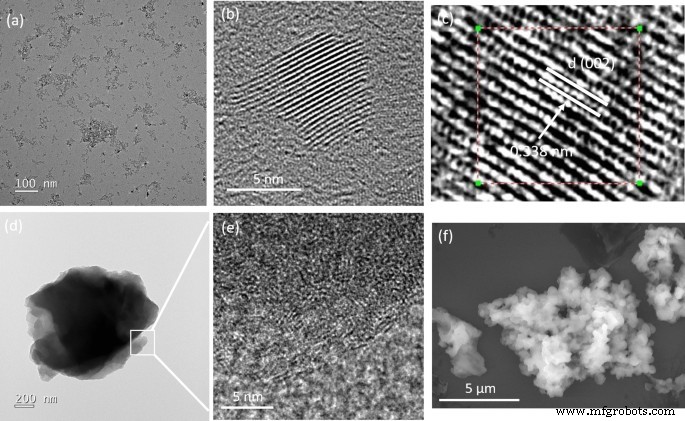
希薄なエタノール溶液から堆積した、調製したままのBGHおよびBMHの代表的な透過型電子顕微鏡写真(TEM)および走査型電子顕微鏡写真(SEM)。 a 、 b BGHの高倍率および低倍率のTEM、 c 0.338 nmの明確な(002)格子間隔を持つBGHの高分解能TEM、 d 低倍率での代表的なBMHのTEM画像 e 図1 d のボックス領域の拡大TEM画像 層状酸化グラフェン内の構造歪みの特徴を示し、 f BMHの代表的なSEM画像
図2は、表1に示す所定の反応条件で当研究室で調製したBMH03(緑のトレース)、BMH01(赤のトレース)、およびBGH01(青のトレース)のXRDパターンを示しています。 43.1°(2θ)。これは、ターボストラティック窒化ホウ素(t-BN)の特徴的な回折パターンです。 26.6°付近を中心とするブロードピークは(002)反射面に由来し、43.1°ブロードピークは六方晶窒化ホウ素(h-BN)によって誘発される(10)反射面に対応します[37]。 BMHのXRDパターンは、2θで約25.4°と42.4°の2つの広い回折パターンによって支配されていました。これらは、それぞれグラファイトの六方晶構造の(002)バンドと(10)バンドの特徴的なパターンです[38]。一般的に示される(10)バンドは、ターボストラティックカーボンの2D反射にも関連しています[39]。さらに、2θ約10.9°にピークがなく、約25.4°に広帯域が出現したのは、グラフェンまたは酸化グラファイトの構造内にドーパントまたは不純物が組み込まれたためです[40、41、42、43、 44,45]。したがって、BMHの主要な構造がドープされた酸化グラフェンであると提案することは合理的です。
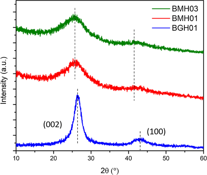
多結晶BMH03(緑のトレース)、BMH01(赤のトレース)、BGH01(青のトレース)、およびBGH01のXRDパターン。カリフォルニアでのBGH01シリーズの幅広いピーク。 26.6°は、h-BNの(002)平面反射と、caでの別のブロードピークを表します。 43.1°は、h-BNの未解決の反射面を表します。 BMHの回折パターンは、約〜25.4°と〜42.4°を中心とする2つの広い反射を示しました。他のBCNOシリーズのXRDパターンは、追加ファイル1:図S3)
に示されています。BGH(グアニジンシリーズ)の構造解析
XPS、FTIR、および固体NMR分光法を実行して、BGHの分子構造をより深く理解しました。 XPSを使用して、B、C、N、およびO元素のコアレベルの電子の存在と、BGH化合物におけるそれぞれの化学結合を確認しました。図3a〜eは、BGHナノディスクの典型的なXPSスペクトルを示しています。 XPS表面元素組成分析によると、BGHには高いBおよびN含有量(それぞれ約40%)が含まれ、CおよびO組成はそれぞれ約8%および13%低くなっています(追加ファイル1:表S3)。 BおよびN組成のほぼ1:1の化学量論は、XRD分析と釣り合っており、私たちの研究室で調製されたBGH01が乱層窒化ホウ素構造で構成されていることを確認しました。すべてのXPSスペクトルは、 R のガウス関数に適合しました。 2 > 0.99。これは、各元素のXPSスペクトルの赤と緑の曲線で表されます。 BGHシリーズの場合、B 1s スペクトルは、B-O結合(189.7 eVの結合エネルギー)とB-N結合(190.7 eVの結合エネルギー)に対応する2つの近似曲線にデコンボリューションされました。 N 1s スペクトルは、397.3eVの結合エネルギーを持つN-B結合と398eVの結合エネルギーを持つB-N-O結合で構成される2つのガウス曲線に適合しました[46]。 N-B結合とB-N–O結合の両方が、OドープhBNの存在またはBNO化合物の形成を示しました[47]。 BGHの合成は大気条件下で行われたため、文献[47]で報告されているように、酸素原子もB-O結合のためにh-BNドメインに組み込まれました。この仮説は、O 1s を分析することによって確認されました。 スペクトル。B-O結合(532 eVでの結合エネルギー)に対応する単一のピークを示しました。この結果は、BGHシリーズがOドープhBNで構成されており、酸素原子の大部分がホウ素原子に結合しているという最初の仮説をさらに裏付けるものです。 C 1s BGHスペクトルの1つは、3つのC種、つまりC-O、C-B、およびC–O–Cにデコンボリューションされ、それぞれ283.7 eV、285 eV、および287eVで結合しました。 BGHシリーズではCの組成が低く、BとNの化学量論が1:1であるため、BGH01の構造はCとOをドープしたh-BNであるとさらに推測しました。提案された構造は、XPSスペクトルで証明されているように、hBNドメイン内でのC-B、B-O、およびB-N–O結合の形成によってサポートされていました(図3)。したがって、800°CでCおよびOドープhBNとして調製されたBGHナノディスクの構造を推定することは合理的です。さらに、粉末XRDと高分解能TEMは、ターボストラティック窒化ホウ素と同様の格子間隔を持つハニカム格子内の共有結合したB、C、N、およびOの形成をサポートしました。この原稿全体を通して、BGH01の構造はBCNOドープhBNと呼ばれます。
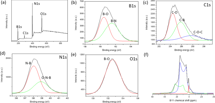
a BGH01のXPS調査スペクトル。 b のコアレベルスペクトル B 1s 、 c C 1s 、 d N 1s 、および e O 1s 。デコンボリューションされた各ピークには、ガウス関数が適用されます。 f 11 BGH01のB固体MASNMRスペクトル。 ( 11 BGH01-LTのB固体MASNMRは、追加ファイル1:図S10)
に示されています。また、他の合成パラメータを一定に保ちながら、BCNOナノ構造の構造光触媒活性に対する焼成温度と時間の影響を調査しました。反応温度を上げ(600°Cから800°C)、反応時間を30分から12時間に増やすと、B-NおよびB-O結合が全体的に増加しました(追加ファイル1:図S6)。対照的に、B-C結合は、反応温度と時間の増加とともに減少します。これは、準安定B-C結合を犠牲にしながら、エネルギー的に安定した六角形のB-NおよびB-O結合の形成を意味します[48]。予想どおり、他のパラメータを一定に保ちながら、ヘキサメチレンテトラミン前駆体(Cソースとして)の比率を増やすと、B-C結合組成が増加します[49]。さまざまな焼成温度、時間、および前駆体比で調製されたBGHの化学結合の進化のより詳細な傾向は、ESIに記載されています(追加ファイル1:図S6およびS7)。
1 11 BソリッドステートMASNMRを使用して、BGHおよびBMHの各B関連結合組成を定量的に分析しました。ここでは、固体MAS 13 を利用した分子レベルでのBCNOの最初の詳細な構造特性を示します。 Cおよび 11 特定のCおよびB関連の結合を収集するためのBNMR。 hBNナノ材料とその欠陥誘起電子特性は大きな関心を集めていますが、hBNエッジの分子構造と欠陥構造はほとんど知られていません[50]。ホウ素関連のナノ材料の構造特性が不足しているのは、固体の 11 の分析が難しいためです。 11 のためのBNMRスペクトル Bは半整数の四重極核( I =3/2)[51、52]。ソリッドステート 11 B NMRは、信号の歪みをもたらす2次四重極結合のため、解釈も困難です。これは、MASNMRを介して部分的にしか平均化できません[53]。さらに、 11 の化学シフトの範囲 B NMRは比較的狭く、さまざまなホウ素種の広く、重なり合い、歪んだピークのピーク割り当ては非常に困難です[54]。この研究では、 11 を実行しました B固体NMRは9.4Tで記録され、スペクトルはトップスピン実線形状分析(SOLA)を使用してデコンボリューションされました。 CP-MAS 11 に従うことによって 文献で報告されているBNMR実験では、四重極結合定数( C )を考慮して、意味のあるB関連の化学結合情報を取得しました。 Q )および電場勾配(EFG)テンソル非対称性(η Q )[51、52、55]。図3fは、ソリッドステートの 11 を示しています。 3つの主要なピークとδを持つBGHのBスピンエコーNMRスペクトル iso それぞれ28.3ppm、20 ppm、1.2ppmを中心にしています。 CP-MASに関する文献研究に基づく 11 窒化ホウ素とその関連構造のBNMR、δのピーク iso 28.3 ppm(緑色のトレースフィット)および C Q 2.85 MHzは、三角形平面BN 2 に対応します。 単一のヒドロキシル基を持つ(OH)種[53,54,55]。ヒドロキシル基または酸素架橋原子が三角形平面のホウ素サイトの周りの窒素原子に置き換わったとき、新しいB種はδ iso 20 ppm(青いトレースフィット)が表示されました。この低い化学シフト信号は、2つのヒドロキシル基または1つのヒドロキシル基と1つの架橋酸素原子(BN(OH) 2 )を持つ別の三角ホウ素サイトに起因する可能性が最も高いです。 またはBNO(OH)サイト)。 δの鋭いピーク iso 1.2 ppm(赤いトレースフィット)は、4配位の四面体Bサイトに対応し、窒素と複数のヒドロキシル基、または架橋酸素原子(52–54)または炭素関連のC-B結合によって配位されている可能性があります[46、52]。 CP-MAS 11 BGH01のBNMR分析により、XPSおよびXRD分析に対応するさまざまなB-N、O-B、およびB-C結合が明らかになりました。さらに、ソリッドステート 11 B NMRは、ほとんどのホウ素が窒素およびBN x として1つ以上のヒドロキシル基と結合していることも明らかにしました。 (OH) 3- x 種族。これらのヒドロキシル化種は、水素結合と静電安定化によって水溶液中でコロイド安定性を提供すると推測されました。また、BN x などの4配位B種の減少も観察されました。 (OH) 4- x またはBN x (O)(OH) 3- x (ここで、Oは架橋酸素です)、反応温度の上昇に伴い、ボロキソールが鳴ります[54、56]。同時に、さまざまな3座標BN x (OH) 3- x アニーリング温度が600°Cから800°Cに上昇すると、種が出現しました(追加ファイル1:図S8およびS10)。この結果は、高温で、ボロキソール環がアンモニアと反応して、さまざまなヒドロキシル化BN x を形成したことを意味します。 (OH) 3- x またはBN x (O)(OH) 2- x 種[55](追加ファイル1:図S10)。
BMHの構造解析
XPSとFTIR、および 11 による精力的な構造分析に基づいています B、および 13 C固体MASNMR、BMHの構造はBCNOをドープした酸化グラフェンであると提案されました。 XPS表面元素分析によると、BMHは75%の炭素種とわずか5%のホウ素関連種で構成されていました(追加ファイル1:表S3)。デコンボリューションされたB 1s スペクトルは、それぞれ191.7および192.4 eVの結合エネルギーでのBCNおよびBN結合を示しました(図4)。 C 1s 種は、C–C結合(sp 2 )に対応する2つの異なる光電子放出シグナルを示しました。 およびsp 3 結合エネルギーが約285.0eVのC–C結合)であり、285.6eVで現れるB-C-N結合からより弱い成分が生じました[57]。 288.0eVおよび288.7eVの他の比較的小さい信号は、C-N 3 によるものでした。 それぞれC =O結合[46、57、58]。酸素化されたC原子(C =O)が酸化グラフェンドメインの端に形成されると推測され、C-N 3 結合はCNの特徴的なピークでした。 N 1s 399.0 eVの結合エネルギーを中心とするXPSスペクトルは、399.2eVの結合でのC-N-Bと400.0eVでのC =Nの結合で構成される2つのガウス曲線に適合させることができます[46、57、59、60]。 XPS分析に基づくと、BMHシリーズのO関連結合の約35%と比較して、BMHシリーズはO関連結合の約10〜25%しか含まれていませんでした。 O 1s 531.2 eVの結合エネルギーを中心とするスペクトルは、C =OおよびCO結合にデコンボリューションできます。これは、酸化グラフェンドメインに起因する可能性があります。 13 C固体CP-MASNMRは、40.2ppmでグラファイトC =C結合の存在をさらに示しました。これは、XRDおよびXPSの結果と一致します[38](図5)。このホワイトペーパーで報告されている各BMHシリーズでは、 13 C NMRは、 C に対応する160および154ppmの炭素種の2つの等モル比を示しました。 α および C β それぞれ、グラファイト状窒化炭素構造[61]に一般的に見られます(図5および追加ファイル1:表S4)。文献手順[34]に従って合成されたバルクCNと比較して、 C のシグネチャー共鳴の化学シフト α および C β ピークはそれぞれ164および156ppmに現れました(図S13)。 CNヘプタジン構造へのホウ素のドーピングは、BMHシリーズとバルクCNの間のこれらのわずかな化学シフトの違いに寄与している可能性があります(追加ファイル1:図S13)。

a Survey XPS spectra of BMH01 and 11 B NMR spectra. Core level spectra of b B1s , c C1s , d N1s , and e O1s 。 Each core spectra were fitted with a black trace, while the red and green traces under the peak were deconvoluted using a Gaussian function. f 11 B solid-state MAS NMR were deconvoluted using SOLA analysis to tricoordinate and tetracoordinate B-sites. The XPS and 11 B solid-state MAS NMR of other BMH series compounds can be found in Additional file 1:Figure S7, S11, and S12)

Solid-state 13 C MAS NMR of a BMH01 and b BMH03
Due to the abundant evidence of the presence of carbon nitride (CN) in the BMH series based on solid-state NMR spectroscopy (Fig. 5), we considered three possibilities of interactions between BCNO-doped graphene oxide and CN in BMH, namely:(i) bulk phase separation, (ii) disordered 2D network, and (iii) layered intercalation [62]. To eliminate the possibility of bulk phase separation, we examined the XRD pattern of CN nitride andBMH as well as a physical mixture of both in a 1:1 mass ratio. We found that the XRD patterns of the mixtures showed only the diffraction pattern of the bulk CN with a slight reduction in the crystallinity compared to the pristine CN diffraction [63] (Additional file 1:Fig S4). Since the XRD pattern of BMH did not possess any diffraction peaks that corresponded to CN, this experimental result confirmed that CN did not form as a bulk-separated domain during the synthesis of BMH (Additional file 1:Fig. S4). We also considered the formation of a disordered 2D network, in which CN and the doped-graphene oxides are bonded on the same 2D plane [62]. Based on a literature report on a CN/graphene oxide 2D matrix, the XRD pattern of a disordered 2D network showed characteristic peaks for both species with a slight peak broadening and a slight peak shifting [64]. However, the XRD pattern of BMH (Fig. 2) did not contain any signature diffractions of CN. A previous study also showed that characteristic peaks of graphene oxide disappeared in a graphitic CN/amorphous CN/graphene oxide composite due to the layer-by-layer interactions [65]. Therefore, it is reasonable to propose that CN is intercalated between the doped-graphene oxide layers. The FTIR spectrum of the selected BGH and BMH series is shown in Additional file 1:Fig. S5.
Boron-related bonding within the BMH series was investigated using 11 B solid-state MAS NMR at 9.4T, and the broad NMR spectrum was deconvoluted using the SOLA analysis showing the presence of both tricoordinate and tetracoordinate boron site. The SOLA analysis yielded four line fittings under the broad 11 B NMR spectrum, which could be assigned as trigonal planar BN2 (OH) or BN2 O at a δiso of 19.8 ppm and a CQ of 2.85 MHz (green fitting). The bay and corner B sites as in B-doped CN appeared at a δ iso of 5 ppm and δ iso of 11 ppm, respectively [61]. These assignments are also commensurate with the formation of CN based on the 13 C NMR and XPS analyses. Compared to the BGH series, the relative composition of the tetracoordinate B(IV) site of BMH was much higher (ca. 55% in the BMH series vs. 3% in the BGH series). However, the tetracoordinate B(IV) species in BMH appeared at a lower chemical shift than those found in BGH (Fig. 3f) and was therefore presumed to be the BN2 (OC)2−x (OH)x species [46]. Notably, the h-BN domain was absent from the BMH series prepared via thermal annealing at a lower temperature (600 °C). However, upon increasing the thermal annealing temperature from 600 °C to 800 °C, the structures of BMH01HT-30 min and BMH01HT-12 h showed a high composition of tetracoordinate BN2 (OH)2 species and the tricoordinate BN3 bonding (Additional file 1:Fig. S11 and S12). The presence of a high composition of tetracoordinate BN2 (OH)2 and BN3 bonding was shared among all the inactive BCNO investigated in this study. Moreover, although BMH01HT-12 h possessed an identical surface elemental composition to that of BGH01, the solid-state 11 B NMR revealed that both compounds possessed significant structural differences, which explained for their differences in photocatalytic activity (Additional file 1:Table S3 and Fig. S12).
In light of the moderate photocatalytic activity of BMH01 and BGH01 (Fig. 7), further synthesis optimization was performed to expand their light absorption spectrum into the visible light region. Previous literature showed that increasing the composition of the hexamethylenetetramine precursor (as a carbon source) could modulate the bandgap and photoluminescence properties of BCNO. Based on these reports, BMH and BGH compounds with a higher ratio of hexamethylenetetramine were prepared accordingly while keeping the other parameters constant. The optimized BCNO with a higher carbon content is denoted as BMH03 and BGH03, in which the molar ratio of carbon source was increased from 0.1 to 0.3. The higher ratio of hexamethylenetetramine precursor yielded BMH03 with a higher composition of the graphitic domain as in sp 2 C = C, and a small peak emerged which corresponded to BCN bonding at 191 eV binding energy (Additional file 1:Fig. S7). The increase in the graphitic sp 2 C = C domain upon increasing the concentration of hexamethylenetetramine is consistent with the role of hexamethylenetetramine as both a C and N source in the synthesis of N-doped graphite [66]. The increased sp 2 C = C graphite bonding in BMH03 was further confirmed via 13 C CP-MAS NMR with the emergence of a more prominent peak centered at 40.2 ppm, which is the signature of graphitic C = C(H) bonding (Additional file 1:Table S4).
Optical Properties
The optical properties of BMH and BGH were investigated using UV–visible absorption and photoluminescence spectroscopy, as shown in Fig. 6. Since BGH and BMH series possessed distinctively different structures, and the optical properties of nanomaterials are highly correlated with their structures, the origin of absorption and luminescence for both series were also found to be different. In this study, BGH01 quasi-spherical nanoparticles showed a featureless UV–vis spectrum. (Fig. 6a, red trace). As the ratio of hexamethylenetetramine increased, the intensity of absorbance peaked at 237 nm for BGH03 and increased with the emergence of an additional broad absorption peak centered around 330 nm (Fig. 6a, blue trace). The origin of the optical properties of BCNO is controversial due to the lack of structural analysis of BCNO nanomaterials. The most widely cited origin of photoluminescence of BCNO is attributed to the formation of B, C, N, and O self-interstitial sites, substitutional impurities, and native point defects within hBN or the graphene matrix [50, 67, 68]. Other possible photoluminescence mechanisms in the BCNO system include the electronic transition from the nitrogen-vacancy (VN ) levels to the carbon impurity levels, the electronic transition between the closed-shell BO − and BO 2− anions, and intrinsic state emission and defect state emission (surface energy traps). The featureless UV absorbance of BGH01 can be attributed to the electronic transition between the valence band (VB) and conduction band (CB) of boron nitride with a bandgap energy of ca. 5.9 eV. The lower energy absorption in BGH03 was a result of the mid-gap absorption from the valence band to the nitrogen-vacancy (VN ) level located approximately 0.7–1.0 eV below the conduction band of hBN.(62) The absorption peak at ca. 330 nm could be attributed to the presence of C-related impurities level located ca. 2–4 eV below the conduction band of hBN [69,70,71]. Under 365 nm excitation, BGH01 produced a broad emission with three bumps located at 412 nm, 445 nm, and 489 nm, respectively. Based on the BGH structural analysis, in which the dominant structure was composed of BN and BO-related bonding, the photoluminescence of the BGH series could be most likely originated from the B-O luminescence centers [24, 47, 72]. The yellow-green emission at 445 and 489 nm could be induced by the transition from the VN level to the carbon-related and oxygen defect levels (2–4 eV) below the conduction band of h-BN [69,70,71]. As the C composition increased in BGH03, the emission wavelength was further red-shifted to 506 nm, consistent with literature reports [73, 74]. The stacked photoluminescence (PL), UV absorbance, and Tauc plot for different BGH series prepared in this study are presented in Additional file 1:Fig. S14.

a Overlay UV absorbance spectra of BMH01, BMH03, BGH01, and BGH03. b Stacked photoluminescence spectra of BMH01, BMH03, BGH01, and BGH03 upon excitation at 365 nm c stacked photoluminescence lifetime decay spectrum of BMH01, BMH03, BGH01, and BGH03 monitored at a predetermined λ max for each sample. Samples were excited with 337 nm laser pulses at 298 K, d Overlay Tauc plot (αhv) 1/2 vs. hv, for BMH01 (black trace) and BMH03 (green trace). The optical band gap is represented by thesolid line, while the interband state for BMH03 is represented by the red dashed line
Based on XRD and various spectroscopic analyses, the structure of BMH was deduced to be dominated by BCNO-doped graphene oxides (Fig. 4, Additional file 1:Fig. S7, and Fig. S12, and Table S4). Thus, the optical properties of the BMH series are hypothesized to be more closely related to the carbon-quantum dot (CD) [75, 76] and doped-graphene oxides systems [44, 77,78,79]. Based on the origin of photoluminescence of the CD and graphene oxides, the optical properties of BMH prepared in this study can be attributed to the intrinsic state emission [76, 80, 81], electron–hole recombination [82, 83], and defective state emission [84]. Intrinsic emission of BMH is speculated to have originated from isolated sp 2 luminescence centers embedded within the sp 3 matrix of the carbonaceous film. The sp 3 matrix of graphene oxides is composed of C–OH, C–O–C, and C = O edge sites, whose energy levels lie between the energy levels of π–π* states of the sp 2 C = C domain, thus giving rise to multiple absorption bands. Both BMH01 and BMH03 possessed a strong UV absorbance band at ca. 240 nm, corresponding to the π to π* transition of C = C within the graphene oxides domain. With the increasing ratio of hexamethylenetetramine in the BMH03 sample, an additional bump at 288 nm emerged, which can be ascribed to the n-π* transition of the C = O and C = N bonds of the oxidized graphitic region [77, 85, 86]. The latter absorbance band at ca. 288 nm was induced by oxygen, nitrogen, and boron defect sites, creating new radiative recombination sites [82, 87,88,89]. Upon photoexcitation at 365 nm, BMH01 and BMH03 exhibited a maximum emission wavelengths at 429 and 447 nm, respectively. The BMH03 sample showed a slight red-shift emission, unlike BMH01 due to the greater extent of graphitization [77, 84] (Additional file 1:Figs. 5 and 6). Both BMH samples revealed a broad and much lower energy emission wavelengths than BGH samples due to lower energy emissive centers arising from O, N, and B defects and surface states. According to the electron–hole recombination mechanism, these photoexcited electrons from each defect state recombine with their corresponding holes in the HOMO, thus yielding a broad photoluminescence emission [90] (Fig. 6b). Interestingly, only BMH01 exhibited a pronounced excitation-dependent photoluminescence as observed in other BCNO [72] and carbon quantum dot systems [76]. The presence of O, and N impurities embedded within the graphene oxides matrix was shown to create a large number of surface emissive traps that corresponded to a diverse energy levels within the bandgap, thus yielding an excitation dependent fluorescence spectra in BMH01 (Additional file 1:Fig. S15). In contrast, the lack of an excitation dependent emission in BMH03 could be explained by the formation of a greater extent of graphitization (C = C) with a concurrent reduction in the surface states population (eg.:C = O) [44].
The bandgap values of BMH and BGH prepared in this study were estimated from Tauc's formulation:(αhν) 2 − hν, where α is the absorbance (Fig. 6d). The bandgap was estimated by extrapolating the photon energy intercept at (αhν) 2 = 0. For the BMH series, the presence of multiple energy levels within the optical bandgap may have originated from the electronic transition from various π-π* (C = C bonds) and n-π* of C = O or other surface groups [76, 82, 87]. As for the BGH series, carbon substituted on boron sites (CB ), nitrogen-vacancy sites (VN ), and interstitial carbon defect levels gave rise to the emergence of interband states between the bandgap of hBN. The presence of multiple energy levels was supported by the photoluminescence spectra of lower energy radiative recombinations [24, 47, 66] (Fig. 6b and Table 2). The BGH03 sample exhibited two large bandgaps at 5.7 eV and 3.8 eV, corresponding to the bandgap of hBN and the transition from the valence band to VN levels, respectively [50, 74] (Additional file 1:Fig. S14).
Long-lived charge carriers that can persist into the microseconds and milliseconds timescales in semiconductor photoelectrodes such as CN photocatalysts, have been proposed as an important parameter in enhancing photocatalytic activity by reducing charge recombinations [91,92,93,94,95]. Time-resolved photoluminescence (TRPL) experiments were conducted to gain insight into the recombination processes of the photogenerated charge carriers of BCNO (Fig. 6c). The µs-PL decay kinetics could be fitted with three exponential decays according to the following equation.
$$I\left( t \right) =I_{1} \exp ( - t/\tau_{1} ) + I_{2} \exp ( - t/\tau_{2} ) + I_{3} \exp \left( {t/\tau_{3} } \right)$$The multiple exponential decays imply that BCNO undergoes complex recombination from both intrinsic and defect states of BCNO [32, 72]. In the equation, I 1 through I 3 are constants with values of emission intensity measured at t =0, andτ 1 through τ 3 are the lifetimes of three channels responsible for the decay, respectively. Through multiexponential fitting of the entire decay curves for the BMH and BGH series, the average lifetimes were calculated to be 1.58, 2.10, 5.18, and 8.14 µs for BGH01, BGH03, BMH01, and BMH03, respectively. The values of I and τ 式で。 1 for the BMH and BGH series are reported in Additional file 1:Table S5. The persistent lifetime of the charge carrier in the BCNO system has been attributed to the presence of shallow traps composed of nitrogen-vacancy (VN ) stabilized by carbon impurities, which were located ca. 0.7 eV-1.0 eV below the conduction band of h-BN [72, 74]. Shallow traps in CN photocatalysts have been attributed to charge separation states with long life-times due to chemical defects [95]. According to works related to prolonged photoluminescence in CN and other nanostructured photoelectrodes [91, 92], the microseconds lifetimes of BCNO-doped graphene oxides and BCNO-doped hBN are associated with the enhanced charged separation within the BCNO domain. The ultralong lifetimes are speculated to be a critical factor in facilitating heterogeneous photocatalysis [91].
Photocatalytic Dye Degradation
As a proof of concept demonstration, the photocatalytic performance of BMH and BGH was evaluated by the photodegradation of methylene blue (MB) under UV–visible light irradiation. Details of the experimental procedure and analysis of the photocatalytic dye degradation are available in the ESI. Figure 7 shows the percent degradation (C /C 0 × 100%) for BMH03 (red line), BMH01 (blue line), BGH01 (green line), BGH03 (orange line), and CN (purple line), where C is the concentration of MB at a time, t and C 0 is the initial concentration MB after the dark equilibrium. According to the Langmuir–Hinshelwood model, ln(C /C 0 ) = kt , where k is the rate constant, the dye degradation rate constant values were calculated to be 2.31 × 10 −3 min −1 for BMH03, 1.52 × 10 −3 min −1 for BGH01, 1.48 × 10 −3 min −1 for BMH01 and 9.38 × 10 −4 min −1 for BGH03. Compared to the state-of-the-art metal-free photocatalyst, BMH03 exhibited a 25% improvement in the photocatalytic dye degradation rates (Fig. 7). This proof-of-concept demonstration warrants a more in-depth investigation of the structure–property relationship of this new metal-free, boron-based photocatalyst.

UV–visible light-induced photocatalytic MB degradation using BGH and BMH. a Photodegradation of MB under UV–visible light (plot of C /C 0 ) and b pseudo-first-order rate reaction kinetics for MB dye using BGH, BMH, and CN as a photocatalyst. c The proposed structures for BGH01, BGH03, BMH01, and BMH03 and their corresponding activities toward photodegradation of MB dye. Each domain is color-coded, i.e., BCNO (orange), h-BN and graphene oxides (blue), tricoordinate boron BN2 (OH) and BO2 (OH) (green), boroxol ring (purple), tetracoordinate B sites (aqua blue), and functional groups or dangling bonds (black).
Based on the detailed structural analysis and the proposed structure in Fig. 7c, the highest photocatalytic activity of BMH03 consisted of BCNO-doped graphene oxides [1, 65]. This ternary metal-free photocatalyst is reported to enhance the photocatalytic performances by increasing the charge separation and migration to the reaction site [1]. Additionally, the incorporation of boron into graphene-based materials [11, 20], CN [21, 96], and carbon nanotubes [97] has also exhibited enhanced performance compared to their pristine material without a dopant due to multiple synergistic effects. The large differences in electronegativity between boron, carbon, and nitrogen (2.04 vs. 2.55 vs. 3.04, respectively) yielded a strongly polarized bonding towards C and N atoms. As a result, a local positive charge was formed on boron that turned boron into a strong acidic defect site for preferential adsorption sites of pollutants [98], O2 [97], HOO
−
and OH
−
[99, 100]. Therefore, electron-rich pollutants such as MB (used as a model reaction) were speculated to preferentially adsorbed onto the electropositive B sites of the BMH photocatalyst. The photocatalytic degradation of MB on BCNO was proposed to undergo an indirect dye degradation mechanism [101]. In the indirect photodegradation mechanism, the photogenerated holes on the surface of BCNO produced highly oxidative hydroxyl radicals and attack the  bond of MB. Meanwhile, photogenerated electrons from the conduction band of BCNO formed highly reducing superoxide radical anion O2
−
species that could directly attack MB. Figure 8 illustrates the proposed mechanism of BCNO in photocatalyzing the degradation of MB
bond of MB. Meanwhile, photogenerated electrons from the conduction band of BCNO formed highly reducing superoxide radical anion O2
−
species that could directly attack MB. Figure 8 illustrates the proposed mechanism of BCNO in photocatalyzing the degradation of MB

Schematic illustration of the proposed mechanism of BCNO in catalyzing the degradation of MB upon light irradiation. The positively charged MB was selectively absorbed on the N2 B-OH sites. The band structures of BMH03 were determined via UPS and Tauc plot (Fig. 6 and Additional file 1:Fig. S17). The redox potentials of O2 /O2 = − 0.13 eV vs ENHE and H2 O/OH = 2.72 eV vs. ENHE are given in black dashed lines as reference
According to the XPS and solid-state NMR analyses, BMH03 possessed a higher composition of graphitic sp 2 C = C with a simultaneous reduction in tetracoordinate B site compared to BMH01 (Additional file 1:Fig. S9 and Table S4) The high composition of tetracoordinate B-sites in BMH01 (33%) also translated to a reduction in the number of boron Lewis acid sites serving as the active catalytic center, which explains the trend of photocatalytic activities among the BMH series [13]. However, the BGH series comprised the C, and O-doped h-BN domain with various tri- and tetracoordinate boron sites. Interestingly, BGH01 annealed at a lower temperature (Table 1, BGH01LT) was found to be inactive but exhibited a high absorbing ability in removing dye from the solution (Additional file 1:Fig. S1 for TEM morphology; Fig. S16 for photodegradation MB UV–vis absorbance).
The previous report also concluded that BCNO was photocatalytically inactive towards dye degradation but exhibited an excellent absorbing ability in removing dye from the solution [102]. The inactive BGH01LT was comprised primarily of tetracoordinate B sites and boron oxides-related bonding. Based on the SOLA analysis and the literature , the tricoordinate boron site with a δ iso of 16.7 ppm was related to boroxol rings (B2 O3 ) [54, 56]. Our results are also supported by other works on the catalytic activation of peroxymonosulfate using amorphous boron [22]. However, the photocatalytic active BGH01, BMH01, and BMH03 possessed a high composition of BN(OH)2 and BN2 OH bonding sites. Our investigation suggested that the photocatalytic activity of BCNO is highly dependent on the local structure of the boron site. While the exact catalytic site and mechanism are yet to be explored, we proposed that BNx (OH)3−x served as one of the BGH and BMH series catalytic sites. For the BMH series, the formation of tetracoordinate B-O sites was detrimental in catalyzing dye degradation due to the reduction in the Lewis acid site. At the same time, the increasing composition of the sp 2 C = C graphitic domain enhanced the photocatalytic activities of the BGH and BMH series.
Conclusions
In summary, BCNO structures and their photocatalytic activities have been presented here and found to be highly dependent on the choice of precursor, precursor ratios, annealing temperatures, and times. In this study, two types of distinctly different BCNO nanostructures were prepared via low-temperature annealing (600 °C–800 °C):(1) BCNO-doped boron nitride and (2) BCNO-doped graphene oxides. Through systematic investigation of using two different nitrogen precursors, crystalline BCNO with a quasi-spherical shape was prepared at 800 °C for 12 hr using guanidine hydrochloride as the nitrogen source. This series of BCNO exhibited moderate photocatalytic activity through the emergence of BN2 (OH) or BN(OH)2 tricoordinate boron serving as the Lewis acidic site. However, BCNO prepared using melamine as the nitrogen source at 600 °C yielded multi-layered sheets with ill-defined shapes. These BCNO-doped graphene oxides layered structures exhibited the highest photocatalytic activity, surpassing the state-of-art metal-free photocatalyst, CN. For the melamine-derived BCNO layered structures, the presence of tricoordinate boron species as BN2 (OH) or BN(OH)2 and a higher composition of graphitic sp 2 C = C were speculated to play an important role in promoting their photocatalytic activity. This study demonstrates the potential of BCNO as a photocatalyst for energy conversion and environmental remediation applications. Further structural optimization on this new B-C-N–O photocatalyst system is expected to facilitate the development of a sustainable catalyst for applications including solar hydrogen fuel production, environmental remediation, electrocatalytic oxygen reduction reaction, and catalytic oxidative dehydrogenation reaction.
データと資料の可用性
All data are fully available without restriction.
略語
- BCNO:
-
Boron carbon oxynitride
- CN:
-
Carbon nitride
- hBN:
-
Hexagonal boron nitride
- TRPL:
-
Time-resolved photoluminescence
- MAS:
-
Magic angle spin
- CP:
-
Cross polarization
- SOLA:
-
Solid line shape analysis
- I :
-
Intensity
- ATR:
-
Attenuated total reflectance
ナノマテリアル