


PEG含有高分子ナノキャリアとの4-チアゾリジンベースの化学療法剤の水分散複合体の増強されたアポトーシス促進効果
要約
目的
4-チアゾリジノン誘導体とPEG含有高分子ナノキャリアとの複合体の水製剤がラット神経膠腫C6細胞に対するアポトーシス促進作用を増強するかどうかを研究すること。
メソッド
4-チアゾリジノン誘導体の抗腫瘍効果のメカニズムを、ラット神経膠腫C6細胞を用いてinvitroで調べた。細胞の出生率、細胞循環パターン、およびアネキシンVの発現を評価し、DNA彗星分析によってDNA損傷を推定しました。高分子ナノキャリアと複合体を形成した4-チアゾリジノン誘導体の新しい水ベースの製剤を利用して、C6細胞に対するアポトーシス促進作用を増強しました。
結果
研究された4-チアゾリジノン誘導体は、G1前段階のこれらの細胞のFACS分析、アネキシンV陽性C6細胞の出現、およびより高いクラスのDNA彗星の数の増加によって確認されるように、ラットグリオーマC6細胞を殺すためにアポトーシスメカニズムを使用します。研究対象の化合物とPEG含有高分子ナノキャリアとの複合体形成により、上記のすべての方法で測定したラット神経膠腫C6細胞のアポトーシス促進効果が大幅に増加しました。
結論
4-チアゾリジノン誘導体とPEG含有高分子ナノキャリアとの複合体形成により、ラット神経膠腫C6細胞における水溶性とアポトーシス促進効果が向上しました。
はじめに
化学療法は多くの腫瘍の主な治療法であり、特に癌がすでに広がっている場合は、化学療法薬が最後の選択肢として選択されます[1]。しかし、重篤な負の副作用は、現在の多くの抗がん剤の臨床効果を低下させます。したがって、副作用が軽減または最小限に抑えられた代替または相乗的な抗がん剤を開発するための継続的かつ決定的な必要性があります[2]。
抗がん剤の水溶性が低いことは、その開発と臨床応用を妨げる一般的で深刻な問題であり、水溶性が低いため、多くの高活性で有望な化学療法剤が除外されています[3]。水不溶性抗がん剤を可溶化するために、界面活性剤と共溶媒が高濃度で使用されます。ただし、これらの物質は有害な副作用を引き起こす可能性もあるため、多くの抗がん剤の使用が制限されます[4]。
医薬品開発の大きな課題は、高活性薬剤、特にその使用を大幅に制限する体内での一般的な毒性を示す抗腫瘍剤の負の副作用を排除または少なくとも軽減することです[5,6,7,8,9 ]。この問題を克服する効果的な方法は、毒性のある抗腫瘍剤が特定の臓器または組織の標的細胞への送達部位で結合することを可能にする薬物の多機能ナノキャリアを使用することです[8、10、11]。ナノ粒子のサイズが小さく、特定の構造が制御され、表面積が大きく、形状が大きいため、他の材料と比較していくつかの利点があります[5、8、9]。ナノ粒子を使用すると、効率を最適化し、負の副作用を最小限に抑え、癌治療を改善することができます[12、13]。したがって、高い一般毒性のために以前に失敗した薬物は、バイオアベイラビリティと制御放出が改善された送達システムに含まれているため、臨床使用の2番目の機会を得ることができます。このような薬物-担体複合体は、血液脳関門や個々の細胞の膜などの自然の障壁をより簡単に通過します。ナノ粒子の表面積が大きいため、特定のベクター生体分子と複合体を形成し、標的臓器への薬物送達を支援し、負の副作用を大幅に低減または完全に排除し、最小有効量を増加させることができます。薬の単回投与中に、薬物作用の有効性を高め、個別化された薬物療法の開発のための新しい刺激を与えます。さらに、ナノ粒子は、特定の薬物の溶解性、安定性、および吸収を改善する分子をカプセル化することができます[11、13、14、15]。薬物送達システムは、血液中の薬物の長期循環、病態生理学的プロセス中に薬物が蓄積する能力、および活性物質の分子を細胞および細胞小器官に効果的に移動させる能力を提供することができる。ナノ粒子は安定していて、一定期間化学構造を維持し、同時に生分解できる必要があります[16、17]。
新しい抗がん剤を評価するには、さまざまな標的ヒトがん細胞株とinvivoでの実験的腫瘍モデルを使用してそれらの細胞毒性活性と効力を測定することが重要です。 化学療法は、腫瘍形成の変化に続く、またはその変化に起因する薬物誘発性細胞死において極めて重要な役割を果たすアポトーシスプロセスの欠陥のために失敗することがよくあります[18、19、20、21]。
多くの悪性細胞はアポトーシス死を回避できるため、新しい抗がん剤の設計と開発には合理的なアプローチを使用する必要があります。新しい抗がん剤を作成するための主な目標は、(1)薬剤作用の独立したメカニズムに影響を与える個々のがん細胞の変異を克服する方法を見つけることです。 (2)独立した経路を同時に標的とすることができる化学療法レジメンを設計する。癌の遺伝学と治療感度との関係をよりよく理解することは、新しい効果的な抗癌剤を開発するための重要な問題です[22]。
以前の研究では、合成4-チアゾリジノン誘導体(Les-3288、Les-3833、およびLes-3882)は、おそらく他の抗がん剤とは異なる作用機序を使用して、ラットC6神経膠腫およびヒトU251神経膠芽腫細胞をinvitroで殺すことを示しました。ドキソルビシン(Dox)に。 Les-3288は、処理された細胞の活性酸素種(ROS)のレベルに有意な影響を与えませんでした[23、24]。これらの強力な抗腫瘍剤は、Doxのものと比較して、腫瘍細胞および動物における毒性作用の測定された生化学的パラメーターによって示されるように、実験動物の体内でより一般的な毒性を示さなかったことを強調する必要があります[7、8]。したがって、抗腫瘍薬と高分子ナノキャリア(PNC)の結合と、安定した水供給システムの形での薬物投与は、遊離型のこれらの物質の作用と比較して、動物の臓器における毒性作用を減らすことができます[ 7、8]。
この研究の目的は、4-チアゾリジノン誘導体とPEG含有PNCの複合体の水ベースの製剤によるinvitroおよびinvivoでのC6系統のラット神経膠腫細胞におけるアポトーシス誘導を研究し、これらの誘導体を使用したアポトーシス誘導を比較することでした。自由形式で。
材料と方法
抗がん剤
複素環式4-チアゾリジノン誘導体(化合物Les-3288およびLes-3833、図1)は、前述のように、ウクライナのダニーロハリツキーリヴィウ国立医科大学の薬学、有機および生物有機化学科で合成されました[25]。
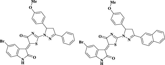
調査した化合物の構造—Les-3288およびLes-3833
細胞培養で使用する前に、これらの化合物をジメチルスルホキシド(DMSO、Arterium、リヴィウ、ウクライナ)に溶解しました。溶液をさらに沸騰水浴中で5分間保持し、作業濃度に達するために蒸留水で希釈した。培地中のDMSOの最終濃度は0.1%未満でした。 Doxは、地元の薬局でウクライナのファイザー(イタリア)の担当者から購入されました。
ポリマーナノキャリア
ドラッグデリバリー用のPNCは、ウクライナのリヴィア工科大学の有機化学科で、前述の方法論を使用して合成されました[26、27]。ポリ(VEP- co </ i> -GMA)-graft-PEGは、次のように次の段階で実行されました。ポリ(VEP- co </ i> -GMA)は、5- tert のラジカル共重合によって合成されました。 ブチルペルオキシ-5-メチル-1-ヘキセン-3-イン(VEP、0.41 g、0.5 mol)(記載された方法[28]で合成された過酸化物モノマー)およびグリシジルメタクリレート(GMA、7.72 g、12.2 mol)(Sigma-Aldrich 、米国)ラジカル開始剤としてアゾイソブチロニトリル(AIBN、0.129 g、0.05 mol)(Merck、ダルムシュタット、ドイツ)を使用した酢酸エチル(7.9 mL)(Merck、ダルムシュタット、ドイツ)中。 65%の最大転化率に達するまで、343Kで重合を行った。ポリ(VEP- co </ i> -GMA)は、ポリ(エチレングリコール)メチルエーテル(mPEG)とサイドエポキシド基の付加反応を介してサイドPEG鎖を結合するためのバックボーンとして使用されました。三フッ化ホウ素エーテル化合物(0.027 mL、0.031 mol)(Sigma-Aldrich、セントルイス、ミズーリ州、米国)を、コポリマー(1.0 g)のジオキサン(20 mL; Merck、ダルムシュタット、ドイツ)の溶液に313Kおよび3時間撹拌した。 mPEG(2.5 mg、3.35 mmol)(Sigma-Aldrich、USA)をジオキサン(15 mL)に溶解し、溶液をバックボーンポリマーの溶液と混合し、313Kで6時間撹拌しました。未反応のmPEGを透析バッグを使用して除去しました。分子量カットオフ(MWCO)の細孔径が6〜8 kDa(Sigma-Aldrich、米国)。結果として生じる櫛のようなポリ(VEP- co </ i> -GMA)-グラフト-PEGを恒量になるまで真空下、室温で乾燥させた。 PNCの構造といくつかの特性を図2に示し、以前に説明しました[29]。
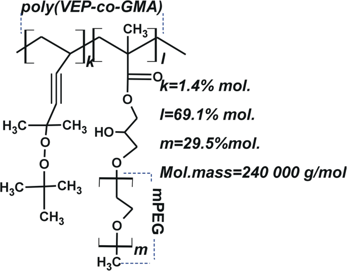
PNCの概略構造— poly(VEP -co- GMA)-グラフト -2- tert の不飽和過酸化物のコポリマーをベースに主鎖を持つPEG -ブチルペルオキシ-2-メチル-5-ヘキセン-3-イン(VEP、「k」で表示)、グリシジルメタクリレート(GMA、「l」で表示)およびポリエチレングリコール(PEG、「m」で表示)の側鎖)
水ベースのPNC溶液を調製するために、0.09gのPNCを0.9mLのDMSOに溶解しました。このPNCの溶液を8.0mLの0.9%NaCl溶液に加えた。次に、溶液を0.5時間撹拌し、10秒間超音波処理しました。複合体の水分散液は、4-チアゾリジノンとPNCのDMSO溶液を次の順序で水に滴下することによって得られました:0.045gのPNCを0.15mLのDMSO(Sigma-Aldrich、USA)に溶解し、0.0015gのLes -3288(またはLes-3833)を0.10mLのDMSOに溶解しました。 PNCと4-チアゾリジノンの溶液を混合し、4.25 mLの1%NaCl水溶液に加え、10秒間超音波処理しました。
この界面活性PNCには、疎水性骨格にグラフトされた親水性PEG鎖が含まれています(図2a)。水溶液中で、両親媒性PNCは、水不溶性化合物(薬物など)を可溶化できるミセルのような構造を形成するか、これらの化合物をナノ粒子の表面に吸着させて、生体適合性を高めます。 4-チアゾリジノンベースの化学療法剤の非常に安定した水分散液を得るために開発された技術は、DMSOの希釈溶液が高分子界面活性剤PNCを含む沈殿剤食塩水に移されるときに発生するナノスケールの薬物粒子の核形成で構成されます。ナノ粒子表面。その結果、凝集と沈降により、複合体が安定し、分散が防止されました。さらに、4-チアゾリジノン誘導体のナノスケール複合体は、周囲の生物学的微小環境との相互作用の可能性から保護されています。この保護により、安定性を失うことなく体内に長く留まり、負の副作用を引き起こすことなく標的組織または臓器に蓄積することができます。したがって、そのような修飾は、抗癌化合物ナノ粒子の水分散液の凝集および沈降安定性を強化します。
PNCのポリマーミセルおよびPNCと複素環式4-チアゾリジノンとの複合体のサイズは、Zetasizer Nano ZS(Malvern Instruments GmbH、シュトゥットガルト、ドイツ)およびDynaPro NanoStar(Wyatt Technology、サンタ)を使用した動的光散乱(DLS)によって測定されました。バーバラ、カリフォルニア、米国)および25°Cでの非侵襲的後方散乱(NIBS)技術を使用した光子相関スペクトルによる。 DLS測定用のサンプルは、上記のように準備し、必要に応じて、pH 6.5〜7.0の再蒸留水で希釈しました。すべてのサンプルに対して3〜5回の測定が行われました(各測定は5サイクルで構成され、測定間の範囲は5分でした)。図3は、PNCのミセル構造とPNCと複素環式4-チアゾリジノン(Les-3833およびLes-3288)との複合体のナノ粒子のサイズ分布をDLSで分析したものです。 PNCミセルおよびPNCコーティングされた4-チアゾリジノンナノ粒子のサイズと形態は、200 kVの加速電圧で透過型電子顕微鏡JEM-200А(JEOL、日本)を使用して研究されました[30、31]。サンプルは上記のように調製し、必要に応じて再蒸留水で希釈しました。サンプルは、基板への均一なコーティングを容易にする超音波分散剤UZDN-1A(Ukrrospribor Ltd.、ウクライナ)を使用して、テストした溶液を基板にスプレーすることによって準備されました。銅グリッド上に堆積された薄いアモルファスカーボン膜が基板として使用された。 DLSおよびTEMの方法で研究されたPNCミセルおよび複素環式4-チアゾリジノン(Les-3288およびLes-3833)との複合体のサイズと形態を図3に示します。
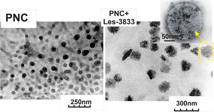
PNC(左)とPNC + Les-3833の複合体(右)の透過型電子顕微鏡(TEM)画像
DLSおよび透過型電子顕微鏡(TEM)研究により、PNCミセルのサイズは50±25 nmであり、PNCと複素環式4-チアゾリジノンLes-3833およびLes-3288との複合体によって形成されたナノ粒子は140±25nmおよび155であることが示されました。それぞれ±30nm 。 PNCと4-チアゾリジノン誘導体との複合体の分散液は非常に安定しており、チアゾリジノンナノ粒子表面に吸着したPNCシェルによる凝集や沈降から保護されています。図4に示すように、水システムに分散したナノ粒子のサイズの変化は、水で複数回希釈した場合、およびPNCと4-チアゾリジノンの複合体の水システムを6か月間エージングした後では無視できます。
>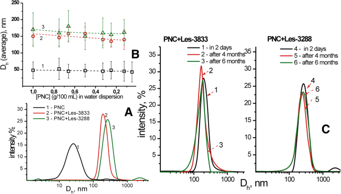
PNCおよび複合体の流体力学的直径のDLS研究( a )および分散希釈( b )に対するPNC-薬物複合体の平均サイズの依存性 ):PNC(1)の水分散液、Les-3833 + PNC(2)、Les-3288 + PNC(3)の複合体、およびLes-3833 + PNC(1–3)とLes-の流体力学的サイズ2日間(1.4)、4か月(2.5)、および6か月(3.6)の保管後の3288 + PNC(4–6)分散液( c )
細胞培養
C6系統のラット神経膠腫細胞は、ウクライナ国立科学アカデミー(キエフ、ウクライナ)の分子生物学および遺伝学研究所の細胞培養コレクションから入手しました。細胞は、10%ウシ胎児血清(Sigma、USA)を添加したダルベッコ改変イーグル培地(DMEM、Sigma、USA)で培養しました。細胞はCO 2 で増殖しました -37°C、5%CO 2 のインキュベーター 、および湿度95%。細胞の再播種は、2〜3日に1回、1:5の比率で実施されました。
研究対象物質の細胞毒性作用の評価
細胞を24ウェルプレートに100,000細胞/ mLの濃度で1ウェルに播種しました。 (Greiner Bio-One North America、Inc。、米国ノースカロライナ州モンロー)。研究中の物質は、細胞播種後に開始された24時間の適応期間の後、0.1、0.5、および1.0μMの濃度で添加されました。細胞計数は、トリパンブルー色素(DV-T10282、Invitrogen、Thermo Fisher Scientific Corp.、米国マサチューセッツ州ウォルサム)を最終濃度0.01%で使用し、細胞懸濁液に添加してから2分後に、血球計算計数チャンバー内で定期的に実施しました。 。死んだ細胞は、原形質膜の損傷のためにこの色素を取り込みました。
フローサイトメトリー
細胞の循環とアポトーシスを研究するために、C6細胞をLes-3288、Les-3833、およびそれらのPNCとの複合体、および遊離型のPNCで処理し、リン酸緩衝生理食塩水(PBS、pH 7.4)で洗浄し、遠心分離によりペレット化しました。 1000 rpmで5分間、4°Cで冷PBSに再懸濁しました(1 mLのPBSあたり200万個の細胞)。次に、穏やかに混合しながら-20°Cに冷却した無水エタノール4mLのアリコートを加えて細胞を固定しました。固定したサンプルは、使用するまで(1週間以内)-20°Cで保存しました。蛍光活性化セルソーティング(FACS)分析では、細胞サンプルを1000 rpmで5分間、4°Cで遠心分離し、上清を捨て、細胞ペレットを1mLのPBSに再懸濁しました。 100マイクロリットルのDNaseフリーRNase(Sigma、USA)をその懸濁液に加え、サンプルを37°Cで30分間インキュベートしました。次に、各サンプルに100μlのヨウ化プロピジウム(PI)(Sigma、USA、1 mg / mL)を添加し、室温で10分間インキュベートしました。最後に、サンプルをプラスチック製のFalconチューブに移し、細胞懸濁液をFACSCaliburフローサイトメーター(BD Biosciences、Mountain View、CA、USA)およびSummit v3.1ソフトウェア(Cytomation、Inc.、Fort Collins、CO、米国)細胞循環とアポトーシスのパラメーターを測定するため。
アネキシンV陽性(アポトーシス)細胞の測定は、FACS分析を使用して実行されました。ラット神経膠腫C6細胞は、0.1 mg / mL、0.5 mg / mL、および1 mg / mLの濃度のPNC、Les-3288、Les-3833、および図に示すようにそれらの高分子コンジュゲートで処理されました。実験の最後に、細胞をトリプシン-EDTA溶液で皿の底から剥がし、PBSで2回洗浄し、アポトーシス検出キット(BD Biosciences、San Jose、CA、USA)を使用してAnnexinV-FITCで染色しました。 、製造元の指示に従って。洗浄した細胞を、1/50容量のFITC結合アネキシンV溶液を含むアネキシンV結合バッファー(BD Pharmingen)で15分間インキュベートしました。次に、サンプルを適切な量のアネキシンV結合バッファーで2倍に希釈し、すぐにフローサイトメーター(Becton Dickinson、USA)のFL1 / FL2(FITC-PI)チャネルで測定しました。アネキシンV-単一陽性細胞はアポトーシスとして分類されました。
DNAコメットアッセイ
DNAコメットアッセイはアルカリ性条件下で実施されました。サンプルあたり75μlの低融点アガロース(0.5%)に1万個の細胞を混合しました。混合後、あらかじめ1.5%の通常の融点のアガロースで覆われたスライドにサンプルをピペットで移しました。サンプルを溶解溶液(2.5 M NaCl、100 mM EDTA、10 mM Tris-base、10%DMSO、1%Triton X-100)中で4°Cで18時間インキュベートしました。 DNAの巻き戻しとアルカリに不安定な損傷の発現を可能にするために、スライドをアルカリ溶液中で室温で20分間インキュベートしました(暗所で実施)。電気泳動(〜74 V / cmで30分間)の場合、スライドを水平チャンバーに移し、1x電気泳動バッファー(0.3 M NaOH、1 mM EDTA、pH> 13)を添加しました。スライドを氷冷した100%メタノールで固定し、乾燥させた。スライドを80μLの1×エチジウムブロマイド(EtBr)で5分間染色した後、冷蒸留水に浸して余分な汚れを取り除きました。 DNA彗星は顕微鏡(Carl Zeiss、ドイツ)を使用して視覚化され、画像はソフトウェア「CASP」(Casplab-1.2.3b2ソフトウェア、CASPlab、ヴロツワフ、ポーランド)を使用して分析されました。サンプルあたり100個の彗星が数えられました。 DNA損傷は、彗星の尾のサイズに応じて5つのレベルの遺伝子毒性に分類されました:0(0–5%の損傷)、1(5–25%の損傷)、2(25–45%の損傷)、3(45–70%の損傷) )、および4(70%以上のダメージ)[32]。損傷指数(DI)は、前述のように計算されました[33]。
メチルグリーン色素を使用したDNAインターカレーションアッセイ
化合物Les-3288およびLes-3833は、メチルグリーンアッセイを使用してDNA分子にインターカレートする能力について調査されました[34]。サーモン精子DNA(10 mg / mL)を、15μLのメチルグリーン溶液(H 2 中1mg / mL)とともに37°Cで1時間インキュベートしました。 O)。化合物を1μg/ mLで添加し、37°Cで暗所で2時間インキュベートしました。サンプルの最終容量の合計は1mLでした。 BioTek 76 883マルチチャネルマイクロフォトメーター(BioTek、Winooski、VT、USA)を使用して、37°Cで2時間サンプルをインキュベートした後、630nmでメチルグリーンの吸収を測定しました。ポジティブコントロールとしてEtBrを使用しました。
データ分析と統計
すべての実験は、各バリアントで3つの緯線を使用して3回繰り返されました。グループの比較には分散分析(ANOVA)を使用しました。細胞周期のさまざまな段階におけるC6ラット神経膠腫細胞の分布のデータは、一元配置分散分析と、それに続くテューキーの事後多重比較検定によって分析されました。すべてのデータは平均±SDとして表されます。 p <0.05の値は統計的に有意であると見なされました。
結果
遊離型および高分子ナノキャリアとの複合体で使用される4-チアゾリジノン誘導体の細胞毒性効果(トリパンブルー排除試験)
トリパンブルー排除試験の結果は、PNCとのLes-3288およびLes-3833複合体の水ベースの形態が、神経膠腫C6細胞(24時間および48時間の治療)に対する毒性が、これらの化合物の遊離型(図5および6)。最高の細胞毒性効果は、PNCとの複合体中のLes-3288およびLes-3833化合物の0.1および0.5μM用量で観察されましたが、これらの化合物の最高用量(1.0μM)では、PNCとの複合体形成の増強が検出されましたLes-3288の場合のみ24時間(図5および6)。
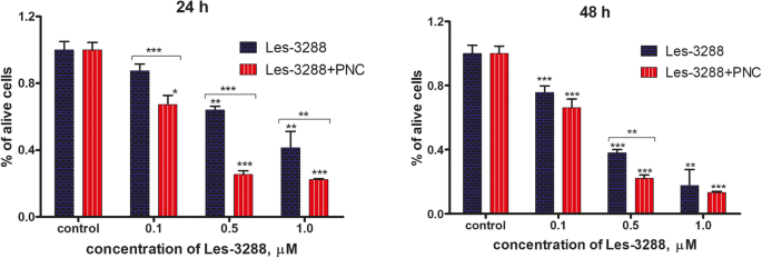
ポリマーナノキャリア( PNC) * P <0.05; ** P <0.01; *** P <0.001 ( コントロールとの違い、100%)
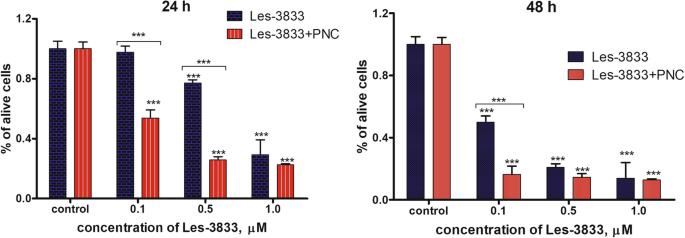
ポリマーナノキャリア( PNC) * P <0.05; ** P <0.01; *** P <0.001(コントロールとの差、100%)
Les-3833およびLes-3288化合物と高分子ナノキャリアの複合体形成は、ラット神経膠腫C6細胞に対するinvitroでのアポトーシス促進活性を増強します
選択した4-チアゾリジノン誘導体の細胞毒性の可能性に対するPNCの調節効果を評価するために、2つの代替アプローチが使用されました。最初に、細胞周期分析を実施して、細胞周期に対する薬物-PNC複合体の影響を調査した。さらに、アネキシンV / PI二重染色を利用して、これらのナノコンポジットによって誘発された細胞死の正確なタイプを区別しました。
低用量(0.1μg/ mLおよび0.5μg/ mL)のLes-3288およびLes-3833化合物は、図7および表1に示すように、細胞周期の進行またはアポトーシスの誘導のいずれにも目に見える影響を与えませんでした。 PNCを含む両方の化合物は、pre-G 1 の細胞数を大幅に増加させました。 フェーズ(Les-3288の場合は4倍、Les-3833の場合は6倍)。これは、アポトーシス促進能の大幅な向上を示しています。遊離型のPNCは細胞周期やアポトーシス誘導に影響を与えなかったことを強調しておく必要があります。したがって、チアゾリジノン-ナノキャリア複合体で治療中のプレG1細胞の大集団の観察された出現は、PNCと実験薬の単純な相乗効果によって説明することはできません。

C6ラット神経膠腫細胞における遊離型および高分子ナノキャリア(PNC)との複合体における4-チアゾリジノン誘導体Les-3288およびLes-3833の細胞周期進行への影響。ヨウ化プロピジウム(PI)染色、フローサイトメトリー。 R2、G1以前; R3、G1; R4、S; R5、G2期。 FL2-H、フローサイトメーターの2番目のチャネル(585/40フィルター)のピーク発光値
観察されたサブG1ピークがアポトーシス細胞で構成されていることを確認するために、アネキシンV / PI染色アッセイの結果を分析しました。低用量(0.1および0.5μg/ mL)および高用量のLes-3288(1.0μg/ mL)でのC6神経膠腫治療中に、ホスファチジルセリン(初期アポトーシスの主なマーカー、FITC結合アネキシンVによって検出)の有意な外在化は観察されませんでした。 、この化合物がアポトーシスの弱い誘導物質であることを示唆している。代わりに、壊死細胞集団の増加(コントロールの1.2%からLes-3288の11.25%、1.0μg/ mL)が見つかりました。これは、Les-3288が少なくとも部分的に壊死細胞死を引き起こすことを示している可能性があります。しかし、Les-3288とPNCの複合体形成は、この薬剤の作用機序を完全に変えました。初期アポトーシス細胞(アネキシンV(+)/ PI(-))と後期アポトーシス細胞(アネキシンV(+)/ PI(+))の数が大幅に増加しましたが、純粋な壊死細胞の総量はコントロール(未処理)細胞のレベル(図8a)。したがって、Les-3288をPNCと結合させると、神経膠腫細胞に対するアポトーシス促進活性が強力に増強されるだけでなく、壊死促進性細胞死の潜在的なメカニズムも低下します。これらの所見は、患者にとって不便な炎症の大規模な誘発のために壊死促進薬の使用が推奨されていないため、臨床診療にとって非常に重要である可能性があります。
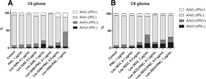
Les-3288のアポトーシス促進活性の比較( a )およびLes-3833( b )ラット神経膠腫C6細胞に対する化合物および高分子ナノキャリア(PNC)とのそれらの複合体。アネキシンV / PI二重染色、フローサイトメトリー。 PI、ヨウ化プロピジウム
驚いたことに、Les-3833 + PNC複合体では同じ効果は観察されませんでした。実際、Les-3833 + PNC複合体のアポトーシス促進活性は、試験したすべての濃度で、遊離型のLes-3288と比較して低かった(図8b)。両方の化合物間のこのような劇的な違いは、それらの化学構造の特異性によって説明される可能性があり、Les-3833の場合、より良い結果を達成するためにポリマーの構造を最適化する必要があると推測されます。
遊離型でPNCと複合体を形成した4-チアゾリジノン誘導体によって引き起こされたラット神経膠腫C6細胞のDNA損傷のコメットアッセイ
アルカリコメットアッセイにより、アルカリに不安定なDNA部位での一本鎖DNA切断を検出できます。得られた結果は、オリーブテールモーメント(OTM)の平均値を使用して推定されました。私たちのデータは、Les-3288、Les-3833、およびPNC(濃度0.5μg/ mL)でのС6ラット神経膠腫細胞の3時間の処理(図9および10)は、重大なDNA損傷を引き起こさなかったことを示しました(OTM =0.7299± 0.1276、OTM =1.466±0.3086、OTM =0.5846±0.1078)、コントロールの未処理細胞と比較(OTM =0.4541±0.07273)。ただし、Les-3833 + PNC複合体(OTM =2.3880±0.2212)を使用した細胞処理は、遊離型のLes-3833を使用した細胞処理よりも重大なDNA損傷を引き起こしました。このような損傷の増加は、Les-3288 + PNC複合体の作用では観察されませんでした(図9および10)。

DNA損傷は、0.5μg/ mLの濃度で使用された実験的抗腫瘍性化合物で3時間処理されたラット神経膠腫C6細胞のオリーブテールモーメントを使用して評価されました。 * P ≤0.05; ** P ≤0.01; *** P ≤0.001。 PNC、高分子ナノキャリア;ドキソルビシン、ドキソルビシン(ポジティブコントロール)
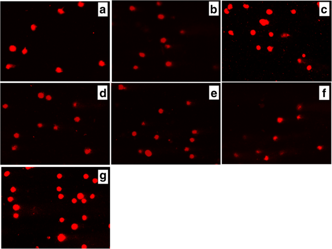
( a で3時間処理した後のラット神経膠腫С6細胞の尾の彗星におけるDNAの存在 )コントロール(未処理の細胞)、Les-3288( b )、Les-3288 + PNC( c )、Les-3833( d )Les-3833 + PNC( e )、PNC( f )およびドキソルビシン( g ), used at a 0.5 μg/mL concentration
An increase of cell incubation time to 6 h (Figs. 11 and 12) caused greater DNA damage in all experimental groups:Les-3288, Les-3288+PNC, Les-3833, Les-3833+PNC, and Dox (positive control). However, the effects of the Les-3288+PNC and Les-3833+PNC complexes appeared to be lower than the effects of the free forms of Les-3288 and Les-3833. It should be noted that the DNA-damaging effect of the complexes of both derivatives with the PNC was less than such effect from these derivatives used in free form.

DNA damage was evaluated using the Olive tail moment in rat glioma C6 cells treated for 6 h with experimental antineoplastic compounds used at a 0.5 μg/mL concentration. * P ≤ 0.05; **P ≤ 0.01; ***P ≤ 0.001. PNC, polymeric nanocarrier; Dox, doxorubicin (positive control)

Presence of DNA in comet tail of rat glioma С6 cells after treatment for 6 h with (a ) control (untreated cells), Les-3288 (b ), Les-3288 + PNC (c ), Les-3833 (d ) Les-3833 + PNC (e ), PNC (f ), and doxorubicin (g ), used at a 0.5 μg/mL concentration
The results were evaluated using the mean value of the TailDNA%. The obtained data show that incubation of С6 rat glioblastoma cells with substance Les-3288, Les-3288+PNC, Les-3833, and PNC (all at concentration of 0.5 μg/mL) during 3 h (Fig. 13) does not lead to significant DNA damage (TailDNA% of 4.157 ± 0.682; TailDNA% of 3.530 ± 1,012, 8.807 ± 0.878; 3.298 ± 0.514, respectively) compared with control (TailDNA% =3.638 ± 0.406), but cell treatment with the Les-3833+PNC complex (TailDNA% =19.53 ± 1.48) causes more significant damage of DNA than the free form of Les-3833. An increase in the incubation time up to 6 h (Fig. 14) in all cases leads to greater damage to the DNA. Again, the level of TailDNA% detected from the action of complexes of both derivatives with the PNC was less than the level of that indicator for the action of these derivatives used in free form.

DNA damage evaluated using the % of DNA in comet tail of C6 rat glioma cells treated with experimental antineoplastic compounds during 3 h at a concentration of 0.5 μg/mL. * P ≤ 0.05; **P ≤ 0.01; ***P ≤ 0.001. PNC, polymeric nanocarrier; Dox, doxorubicin (positive control)

DNA damage evaluated using the % of DNA in comet tail of C6 rat glioma cells treated with experimental antineoplastic compounds during 6 h at a concentration of 0.5 μg/mL. * P ≤ 0.05; **P ≤ 0.01; ***P ≤ 0.001. PNC, polymeric nanocarrier; Dox, doxorubicin (positive control)
We also used an alternative, five-category classification system to classify DNA migration and calculated the DI. Table 2 summarizes the percentages of rat glioma C6 cells for each level of genotoxicity during 3 h of treatment. In the control (untreated cells), higher percentages of cells in levels 0 (81.3%) and 1 (18.6%) were detected compared to these levels in Les-3288 and PNC-treated cells. Complexation of Les-3288 with the PNC did not lead to a big change in the distribution pattern of the DNA comets. In contrast, Les-3833-treated cells showed a lower percentage of cells in 0 level (38.6%) and a higher percentage of cells in level 1 (60.0%), while Les-3833 + PNC-treated cells demonstrated a significantly lower percentage of cells in level 0 (8.0%) and much higher percentages of cells with levels 1 (76.0%), 2 (13.3%), and 3 (2.6%). The integrative indicator of DNA damage (DI) in the case of application of both Les-3288 and its complex with the PNC did not differ significantly from that for control or using free PNC, while the DI from using both Les-3833 and its complex with the PNC was even higher than the DI from using Dox.
<図>During this time interval, Les-3833 + PNC-treated cells demonstrated a higher percentage of cells with level 4 (5.3%) DNA damage than cells treated with Les-3288 and the other experimental compounds, but the percentages of cells in levels 2 and 3 were lower (16.0% and 12.0%, respectively) than those from treatment with other compounds (Table 2). Table 3 summarizes the percentages of rat glioma C6 cells with DNA damage after 6 h of treatment. In general, a tendency for the appearance of DNA comets of higher classes was detected when complexes of Les-3833 and Les-3288 were used, compared with a spectrum of DNA comet classes induced by free forms of these agents (Table 3). However, the integrative indicator of DNA damage (DI) calculated for the action of complexes of Les-3833 and Les-3288 was found to be lower than such an indicator for the action of free forms of these agents. A possible explanation could be a very high DI level at 6 h action of studied agents that is even higher than such indicator found for the action of Dox (Table 3). Thus, we observed time dependence of DNA damage caused by complexes of Les-3833 and Les-3288:at 3 h of treatment, there was an increase of the DI in the case of action of Les-3833 complexes, compared to the action of the free form of Les-3833; however, at 6 h of treatment, there was a decrease in DNA damage for the action of complexes of both Les-3833 and Les-3288, compared to such damage observed for the action of the free forms of these agents. There was no significant time dependence for the action of the PNC, while a significant time-dependent (from 3 to 6 h) increase in the DI was observed for the action of Dox (from 41.2 + 0.40 to 185.2 + 0.23).
<図>DNA Intercalation by 4-Thiazolidinone Derivatives—Les-3288 and Les-3833
Les-3288 and Les-3833 compounds demonstrated a certain capability for intercalating DNA molecules with approximately 15% and 10%, respectively, of replacement of methyl green dye. EtBr, a well-known DNA intercalating agent, was used as a positive control, and it was shown to replace the methyl green dye much more efficiently (70%) (Fig. 15). Thus, Les-3288 and Les-3833 are not capable of effectively intercalating in between two complementary base pairs in double-stranded DNA.

Replacement of methyl green dye intercalated into the DNA of salmon sperm by Les-3288 and Les-3833 compounds (1.0 μg/mL) and ethidium bromide (EtBr) used as a positive control
Discussion
There are two major problems that impede increasing the efficiency of treatment for cancer patients:(1) non-addressed actions of many anticancer drugs that lead to general toxicity and severe negative side effects due to damage of normal tissues and organs; (2) rapid (6–12 months) development of resistance of tumor cells to applied anticancer drugs that leads to a loss of efficacy of drug action. In addition, there is a third, technical problem of poor water solubility of many anticancer drugs. However, binding these drugs with specific carriers of the nanoscale size can help to solve the solubility problem.
In this study, we addressed the first and third of the above-mentioned problems by (a) selecting novel synthetic substances (4-thiazolidinone derivatives:Les-3288 and Les-3833) that possess both high anticancer activity and less general toxicity in tumor-bearing animals [7, 8, 23] and (b) applying a novel drug delivery platform that provides stable water-based forms of the complexes of the water-insoluble 4-thiazolidinone derivatives with a PEGylated polymer.
The 4-thiazolidinone derivatives—Les-3288 and Les-3833—are heterocyclic compounds with molecular mass of 400–600 Da. These and other related derivatives were synthesized at Lviv National Medical University (Ukraine), and most of them have been tested under the Developmental Therapeutic Program at the National Cancer Institute in Bethesda, MA, (USA) [35,36,37,38]. Based on these evaluations of their antineoplastic activity, the most promising substances were selected for further study of the mechanisms of their cytotoxic action and anticancer effects [25]. Les-3833 demonstrated high toxicity towards B16F10, WM793, and SK-Mel-28 melanoma cells, and against human lung A549, breast MCF-7, colon HCT116, and ovarian SKOV3 cancer cells and leukemia cells (L1210, Jurkat, HL-60 lines) [23, 25, 39]. Les-3288 and Les-3833 showed high toxicity against mammalian glioma cells (C6, U251, U373 lines) [23, 24].
Earlier, we found that Les-3288 and Les-3833 were the most toxic among the other studied 4-thiazolidinone derivatives towards rat glioma C6 cells and human glioblastoma U251 cells [23, 24], although their application for cell treatment was very complicated due to their absolute insolubility in water. Only using DMSO with additional heating was effective for preparing a soluble form of the derivatives (see the “Materials and Methods” section). Thus, it was reasonable to search for a way of improving the delivery of these derivatives to target cells, and we used a synthetic PNC to accomplish this task. The complexation of the studied derivatives of 4-thiazolidinone with the applied PNC also enhanced the effectiveness of their antineoplastic action. The mechanisms of the pro-apoptotic action of these compounds were demonstrated using western blot and FACS analyses [23, 24]. It should be noted that in healthy experimental animals, Les-3288 and Les-3833 produce much fewer negative side effects such as cardiotoxicity [40], hepatotoxicity [8], and nephrotoxicity [7] than the widely used anticancer chemotherapeutic agent Dox.
In a previous study, we showed that Les-3288 did not induce ROS production during its pro-apoptotic action in vitro and effects in experimental laboratory animals [23], while the action of Les-3833 led to such production; however, it was less than that of Dox. ROS are considered to be the main effectors of negative side effects of anticancer drugs, particularly Dox [41, 42]. Thus, the Les-3288 and Les-3833 compounds could be prospective anticancer drugs because they possess antitumor activity, but do not demonstrate general toxicity at the level characteristic for effective anticancer medicines such as Dox.
A great impediment to promoting Les-3288 and Les-3833 as anticancer drugs is their poor water solubility. This problem also exists for other anticancer medicines, such as taxols [2, 4]. For paclitaxel, this problem was solved by using a special oil for preparing the medicinal formulation [2, 4, 43]. However, specific delivery platforms are considered to be more promising for creating “smart” drugs that possess effective action in the organism [3, 13]. Specific functional polymer surfactants with block, comb-like, and block/branched architectures, including PEG-containing ones, as well as derived water forms have been developed and proposed for application as carriers for the delivery of Dox [26, 27] and the metal chemotherapeutic agent KP-1019 [10] at the Department of Organic Chemistry of Lviv Polytechnic National University.
Since the 4-thiazolidinone derivatives are water-insoluble compounds, DMSO was used to prepare their water-soluble forms. To avoid the negative consequences of using a toxic solvent like DMSO, we complexed these derivatives with the above-noted PNC. Highly dispersed and stable nanoscale water dispersions of these substances were obtained and applied for treatment of rat glioma C6 cells.
The use of complexes of 4-thiazolidinone derivatives with the PNC led to reduced general toxicity of these compounds in experimental animals. Earlier, we found that when experimental antitumor agents (Les-3288, Les-3833, Les-3882) were applied in a complex with a synthetic polymeric carrier, their action was accompanied by much smaller changes in the biochemical parameters in blood serum that are characteristic for cardiotoxicity [40], hepatotoxicity [8], and nephrotoxicity [7], compared to those for Dox. Thus, complexation of these antitumor agents with the PNC and their application in the form of water-soluble stable delivery systems reduces their toxic effects in experimental animals, compared with the action of these substances in a free form [44]. Several systems for paclitaxel delivery have been proposed including polymeric nanoparticles, lipid-based formulations, polymer conjugates, inorganic nanoparticles, carbon nanotubes, nanocrystals, and cyclodextrin nanoparticles [43].
Thus, complexation of Les-3288 with the PNC significantly increased the pro-apoptotic activity of this experimental drug, thereby allowing us to advance it to pre-clinical studies on animal tumor models. Complexation of Les-3833 with the same type of nanocarrier was less efficient, and thus another type of polymer should be used.
The developed water-based forms of the complexes, as well as the free derivatives, were applied for treatment of rat glioma C6 cells. It was found that these water-based forms of the complexes of the PNC with the 4-thiazolidinone derivatives—Les-3288 and Les-3833—enhanced the pro-apoptotic action of these compounds. In another study, we demonstrated that the induction of apoptosis by Les-3288 was not accompanied by ROS induction, opposite to the action of Dox and Les-3833 [23]. Thus, high antitumor activity of Les-3288 together with its low general toxicity when targeting the malignancy in NK/Ly lymphoma-bearing mice suggest great potential for this experimental anticancer compound. The presented data also predict the potential usefulness of Les-3288 and Les-3833 compounds complexed with the PNC for glioma treatment.
Cell division, differentiation, and death are principal physiological processes that regulate tissue homeostasis in multicellular organisms. A disruption of genomic integrity and impaired regulation of cell death may both lead to uncontrolled cell growth. A compromised cell death process can also promote genomic instability. It is becoming clear that dysregulation of the cell cycle and cell death processes plays a pivotal role in the development of major disorders such as cancer, cardiovascular disease, infection, inflammation, and neurodegenerative diseases [45].
We have found that some of the 4-thiazolidinone derivatives, namely, Les-3288 and Les-3833, were even more effective than Dox in the induction of apoptosis in rat C6 glioma and human U251 glioblastoma cells in vitro [23, 24]. It should be noted that brain tumors belong to malignancies that are the most resistant to applied chemotherapies, and survival rates in patients with these tumors are extremely low [44].
It has been shown that the complexation with this PNC enhanced the pro-apoptotic action of 4-thiazolidinone derivatives (Les-3288 and Les-3833) [44]. In another study [23], we demonstrated that the induction of apoptosis by Les-3288 was not accompanied by ROS induction, opposite to the action of Dox and partially opposite to that of Les-3833. These data could explain the lower general toxicity of Les-3288 compared with that of Dox [23, 24].
The results of FACS analysis are in agreement with the suggestion that an enhancement of the pro-apoptotic action of the studied 4-thiazolidinone derivatives is due to their complexation with a novel PNC. Such complexation of Les-3288 and Les-3833 increased the ratio of pre-G1 cells in the population of treated rat glioma C6 cells, decreased the ratio of G1 cells, and increased the action of G2/M cells. These data suggest an enhancement of both cytotoxic action (apoptosis) and cytostatic action (block in G2/M phase of cell cycle) of the studied derivatives complexed with the PNC. In general, the apoptosis mechanism of glioma cell killing by the Les-3288 + PNC complex and the Les-3833 + PNC complex was demonstrated by the results of FACS analysis of the appearance of Annexin V single-positive rat glioma C6 cells.
Involvement of the apoptosis mechanisms in the action of the studied 4-thiazolidinone derivatives was also confirmed by the results of the DNA comet assay. It was found that the complexation of Les-3833 with the PNC led to a decrease (compared to free Les-3833) in the ratio of rat glioma C6 cells in “0” DNA comets and sіmultaneous increase in the ratio of “1,” “2,” and “3” comets (3-h treatment of cells). At 6 h of treatment by the Les-3833+PNC complex, there was a large increase (compared to free Les-3833) in the ratio of “1” comets, an increase in the ratio of cells with the most expressed DNA damage (“4” comets), and a decrease in the ratios of “2” and “3” comets. It should be noted that the DNA damaging effects of Les-3288 and Les-3833 measured at 6 h was higher than such effects of complexes of these derivatives with the PNC (see Table 3). A possible explanation here could be a very high level of the DI at 6 h action of studied agents that is even higher than the level of that indicator found for the action of Dox (Table 3). There was no time dependence in the action of the PNC, while a significant time-dependent (from 3 to 6 h) increase (from 41.2 + 0.40 to 185.2 + 0.23) in the DI was detected for the action of Dox.
Thus, a complexation of 4-thiazolidinone derivatives—Les-3288 and Les-3833—with a PNC enhanced the cytotoxic effect of these compounds towards rat glioma C6 cells, and that effect was achieved through the apoptosis mechanisms including DNA damage observed as DNA comets in the electrophoresed cells.
We used the DNA intercalation test to investigate the direct targeting of DNA by the 4-thiazolidinone derivatives and revealed only weak replacement of methyl green dye (15% and 10% replacement for Les-3288 and Les-3833, respectively) that intercalated into the DNA of salmon sperm, compared to the replacement by EtBr, used as a positive control (70%).
Thus, conducting the cytotoxicity experiments in vitro with the 4-thiazolidinone derivatives—Les-3288 and Les-3833—was more convenient due to using the water-based forms of the 4-thiazolidinone derivatives—Les-3288 and Les-3833—complexed with the novel PNC. Besides, we have demonstrated that the novel synthetic 4-thiazolidinone derivatives (Les-3833 and Les-3288) possessed treatment effects i n vivo towards NK/Ly lymphoma grafted to BALB/C mice, and those effects were comparable to such effects of Dox. At the same time, the action of these derivatives led to much fewer negative side effects measured as changes in the number of erythrocytes, neutrophils, and lymphocytes in the animals’ blood and the activity of aspartate and alanine aminotransferases in their blood serum, compared with the action of Dox.
結論
The complexation of Les-3288 and Les-3833 with the PEG-containing PNC enhanced their cytotoxic action towards rat glioma C6 cells. The cytotoxic action was realized by apoptotic mechanisms and confirmed by FACS analysis as the presence of the pre-G1 fraction in the rat glioma C6 cells and of Annexin V-single-positive cells. DNA comet analysis revealed single-strand brakes in the nuclear DNA of treated glioma C6 cells that probably was not caused by the intercalation of the studied compounds into the DNA molecule. The Les-3288 and Les-3833 stabilized by the amphiphilic PEG-containing PNC resulted in the water-based forms and provided enhancement of their antitumor effect in vitro in comparison with the free form of the drugs.
略語
- DMSO:
-
ジメチルスルホキシド
- Dox:
-
ドキソルビシン
- EtBr:
-
Ethidium bromide
- FACS:
-
Fluorescence-activated cell sorting
- ROS:
-
活性酸素種
- PBS:
-
リン酸緩衝生理食塩水
- PEG:
-
ポリエチレングリコール
- PI:
-
ヨウ化プロピジウム
- PNC:
-
Polymeric nanocarrier
- SEM:
-
走査型電子顕微鏡
- TEM:
-
透過型電子顕微鏡
- VEP:
-
2-Tret-butylperoxy-2-methyl-5-hexen-3-in
ナノマテリアル
- Zn-Al層状複水酸化物による光触媒的に強化されたカチオン染料の除去
- フォトニック結晶に基づくハイブリッド液晶セルにおける強化された非線形光学効果
- 多機能GaN / Feナノ粒子による内皮細胞の標的化
- 光触媒性能が向上した新規Bi4Ti3O12 / Ag3PO4ヘテロ接合光触媒
- 光触媒活性が強化されたAgナノ粒子/ BiV1-xMoxO4の相乗効果
- Er3 + -Yb3 + -Li +トリドープTiO2のアップコンバージョン材料によるペロブスカイト太陽電池の電力変換効率の向上
- 全炭素3Dナノ濾過膜による水からの抗生物質の除去
- Al / PTFEナノラミネートとの統合に基づく強化されたエネルギー性能
- ポリ(3-ヘキシルチオフェン)中の金属硫化物ナノ結晶のその場成長:[6,6]-光電流が強化された反転ハイブリッド太陽電池用のフェニルC61-ブチル酸メチルエステル膜
- トランスフェリン受容体による細胞内在化が増強されたパクリタキセルベースの標的脂質ナノ粒子の抗増殖およびアポトーシス誘発能—白血病細胞での研究
- 高速セレニル化アプローチによるSb2S3平面ヘテロ接合太陽電池の強化された光起電力特性