


生物医学、ドラッグデリバリーおよびイメージングアプリケーションのための磁気機能化ナノ粒子
要約
医学は、高い有効性と費用効果が必要な病気の新しく改善された治療法を常に探しており、そのような新しい治療法を発見するための科学的研究に大きな需要を生み出しています。治療の重要な側面の1つは、病気だけを対象とし、体の別の健康な部分に害を及ぼさないようにする能力です。このため、金属ナノ粒子は、医用画像処理、抗菌および抗ウイルス用途を含む、それらの可能な医療用途について広く研究されてきました。超常磁性金属ナノ粒子は、磁場で体の周りに向けたり、磁気インプラントに向けたりできる特性を備えています。これにより、さまざまなバイオカーゴをナノ粒子に結合させ、体内での治療に向けることができます。ここでは、単一金属ナノ粒子、機能化金属ナノ粒子、Fe 3 のコアを使用したコアシェル金属ナノ粒子など、さまざまな金属ナノ粒子の現在の生物医学的応用について報告します。 O 4 これらのコアシェルナノ粒子の合成方法と同様に。
はじめに
金属ナノ材料は、医学の未来への重要な入り口を表しています。医学における金属ナノ粒子の長期的な安全性についてはまだ多くのことがわかっていませんが[1]、これらの粒子は、in vivoでの部位特異的イメージング[2,3,4]、癌の検出など、さまざまな生物医学的用途ですでにその位置を占めています。 [5、6]、癌治療[7,8,9,10]、神経変性疾患治療[11,12,13]、HIV / AIDS療法[14,15,16]、眼疾患治療[17,18,19 ]、および呼吸器疾患治療[20、21]。図1は、医療におけるナノ粒子の現在のさまざまな用途を示しています。ナノ医療の最近の進歩にもかかわらず、ナノ治療の方法には依然として多くの障害があります。たとえば、多くのナノ粒子合成法は両方のサイズの範囲を生み出すため、簡単に再現可能な結果を生み出す合成経路を達成するのは難しい場合があります[22 、23,24]およびナノ粒子の形状[25,26,27,28]および/または経済的に実行可能にするのに十分な量のナノ材料を生成しない[29]。もう1つの重要な要因は、研究分野が比較的新しいため、一定期間にわたる一部のナノ粒子の長期毒性については比較的不明であるということです[30、31、32]。金属ナノ粒子の多くの可能な用途の中には、ドラッグデリバリーの分野があります[33、34]。ナノ粒子は表面積が大きいため[35]、大量の薬物やその他の医療貨物を運ぶことができる能力を備えています[36]。単一金属ナノ粒子の代替は、シェル材料に代替の特性を有するナノ粒子にコアを組み込むことであり、これの一例は、磁気コアを組み込むことである。まだ存在している課題の1つは、コアシェルナノ粒子の合成です。ナノ粒子を合成する方法はたくさんありますが[37]、コアシェルナノ粒子を合成しようとすると新しい課題が発生します[38]。

医学におけるナノ粒子の現在の使用のいくつか
このレビューでは、最初に、Fe 3 のコアの使用に焦点を当てたコアシェルナノ粒子の生成に現在使用されている方法のいくつかに焦点を当てます。 O 4 金または銀のコーティング。次に、単一金属ナノ粒子の現在の生物医学的応用、それらの限界、および磁気コアの応用でそれらを克服する方法を検討します。
コアシェルナノ粒子の合成
金属ナノ粒子の合成方法は、長年にわたって知られており、例えば、Stevenson etal。 HAuCl 4 の還元による金ナノ粒子の合成を発表 1951年[39]。それ以来、ガス堆積[40]、ゾル-ゲル[41]、エアロゾル/気相[42]など、ナノ粒子合成にはさまざまなルートがあります。しかし、コアシェル構造からなる金属ナノ粒子を合成しようとすると、新しい課題が発生します。この場合、1つの金属がコアを形成し、2番目の金属がシェルを形成します。たとえば、Fe粒子は水中で分解しますが、HAuCl 4 強力な酸化剤です[38]。さらに説明するそのような例の1つは、Fe 3 を使用することです。 O 4 (酸化鉄)コアとコーティングシェルとしての金。このようなコアシェル金属ナノ粒子の調製において、最大の問題の2つは、コーティングの速度を制御し、コーティングの均一性を制御して、すべて非常に類似した形状とサイズのナノ粒子の溶液を作成しようとすることです[43]。酸化鉄コアへの金または銀のコーティングは、2つの主要なカテゴリに分類できます。鉄への金/銀の直接コーティング[44]、または中間層を使用して金と鉄層の間の接着剤として機能する[45]。ここでは、前者のカテゴリについて説明します。次のテキストでは、金と銀でコーティングされたFe 3 を合成するために考案されたいくつかの方法について説明します。 O 4 ナノ粒子。
逆ミセル合成
金属ナノ粒子を合成するための一般的なルートは、逆ミセル法を使用することであり、マイクロエマルジョンルートと呼ばれることもあります[46]。この方法は、ロジウム、白金、およびパラジウムのナノ粒子のコロイド溶液が最初に合成された1980年代に最初に導入されました[47]。
ミセルは、疎水性および親水性の構成部分を持つ分子が水相または疎水性相のいずれかと接触したときに形成されます[48]。ミセルは、親水性部分が水相と接触し、疎水性成分が疎水性相に面するように組織化されます[49]。本質的に、回転楕円体は内部シールド相で形成され、さらに貨物を収容することができます[43、50、51、52]。
マイクロエマルジョンルートにはさまざまなアプローチがあり、これらには油中水(w / o)[53]および超臨界中水CO 2 が含まれます。 (w / sc-CO 2 )[54]。水が炭化水素ベースの連続相に分散し[53]、熱力学的に駆動される界面活性剤の自己組織化により、w / oエマルションが発生し、逆ミセルが生成されます。球状ミセルが最も一般的な形状です[43]。この混合物に追加された極性またはイオン性材料はミセル内で区画化され、ブラウン運動によってミセル膜が互いに収縮するとナノ粒子が形成されます[55]。 w / sc-CO 2 エマルジョンには、液体(CO 2 )超臨界状態にある、つまり臨界圧力と温度の両方を超えている[56]。この方法は、有毒な有機溶媒を必要としないため、ナノ粒子合成へのより「グリーン」なアプローチであるため、特に興味深いものです。また、圧力を下げて流体をCO 2 として放出するだけで、製品を回収するのも簡単です。 ガス[57]。
逆ミセル経路は、金属ナノ粒子の合成から以前に合成されたナノ粒子のコーティングに適応されています[58]。最初の金でコーティングされた酸化鉄(Au-Fe 3 O 4 )逆ミセルで合成されたナノ粒子は、ほぼ20年前に行われた[59]。このAu-Fe 3 の合成 O 4 ナノ粒子はH 2 を使用して行われました 水素化ホウ素ナトリウム(NaBH 4 )でミセルを生成するO / CTAB(セチルトリメチルアンモニウムブロミド)システム )還元剤として、塩化金(HAuCl 4 )鉄心に。この合成により、平均サイズが12nmのナノ粒子分散液が生成されました。これはAu-Fe 3 の最初の生産であるため O 4 マイクロエマルジョンを使用するNPには、さまざまなAu-Fe 3 があります。 O 4 発見されたNP合成経路[46、60、61、62、63]。図2は、逆ミセル経路を使用してナノ粒子がどのように形成されるかを一般的に表したものです。
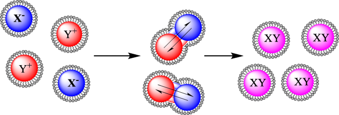
塩を含む逆ミセルの相互作用の一般的な表現は、反応して金属ナノ粒子を形成します
リンら。 Fe 3 をコーティングするためのわずかに変更された方法を公開しました O 4 逆ミセル法を使用して金で[60]。合成では、界面活性剤としてCTABを使用して逆ミセルを形成するシステムも使用しますが、共界面活性剤として1-ブタノール、油相としてオクタンを使用し、NaBH 4 HAuCl 4 を減らす 酸化鉄ナノ粒子の表面に。報告されたコーティングされた粒子の光学的結果は、UV / visスペクトルの吸光度ピークが金コロイド(526 nm)からAu-Fe 3 にシフトしていることを示しています。 O 4 (555 nm)。コーティングされた粒子のTEM結果は、5〜15 nmのサイズ分布を示し、平均サイズは10nmでした。この方法は、パナらによって繰り返されました。 5〜35nmサイズのAu-Fe 3 のわずかに大きいサイズ分布 O 4 ナノ粒子[63]。さらに、非常に類似したシステムがSeip etalによって採用されています。 HAuCl 4 を減らすためにヒドラジンを使用することを除いて [64]。
Fe 3 のコーティング O 4 ナノ粒子は金だけに限定されません。 Lopez Perez etal。シクロヘキサン/ Brij-97(共界面活性剤)とFeSO 4 の鉄塩を含む水相を含むシステムを使用した酸化鉄ナノ粒子の合成について報告されています。 .7H 2 OおよびFeCl 3 .6H 2 O [65]。このシステムは、銀[58]と金[46]の両方でコーティングされており、13nmの粒子を生成します。別の方法がTamerらによって報告されています。 Au-Fe 3 の合成用 O 4 ナノ粒子[62]。この方法では、NaOH中の鉄塩の共沈を採用し、次にHClO 4 で洗浄しました。 酸化されたFe 3 を生成する O 4 ナノ粒子。 Fe 3 への金のコーティング O 4 NPは、HAuCl 4 の還元を介して発生しました CTABミセルによってシステムに供給されるNaOHによって。 Au-Fe 3 O 4 NPは23.5nmの平均サイズで生成されました。特性評価後、粒子はさまざまな官能基で修飾されて自己組織化単分子膜(SAM)を形成し、さらに Escherichia coli の捕捉と検出に使用されました。 。
逆ミセル合成の修正版は、Zhangらによって行われています。鉄ナノ粒子を金でコーティングするための開始剤としてレーザーを使用することを含む[66]。このプロセスでは、CTABミセルにカプセル化された鉄ナノ粒子、水中の金ナノ粉末、およびオクタンの反応混合物を作成し、反応を激しく攪拌しながらパルスレーザーを照射します。レーザー照射は、金ナノ粒子の熱分解を促進します。金の原子とクラスターが鉄のナノ粒子の周りに形成され、金でコーティングされた鉄のナノ粒子を形成します。この方法で合成されたAu-Feナノ粒子のTEM結果は、平均サイズが18 nm、サイズ分布が±36nmでした。
熱合成
金のシェル-鉄コアナノ粒子合成のさまざまな方法の中には、反応が沸点を超えるまで反応混合物を加熱し[67]、時には還流する[68、69]という熱経路があります。このタイプの合成には、主に2つのカテゴリがあります。水熱(水ベースの溶媒)[70、71]とソルボサーマル(有機ベースの溶媒)[68、72]です。熱経路を介して金属ナノ粒子を合成するための多くの技術がありますが[73,74,75,76,77,78]、ワンポット反応でコアの合成と金のコーティングを達成することはできません[68、 69、72、74、77、79、80、81]、場合によってはFe 3 O 4 コアは逆ミセル経路[70]またはコロイド経路[78]を介して合成され、次に粒子は水熱またはソルボサーマル技術[70、76、78]を使用してコーティングされます。これらの合成方法で使用されるさまざまな溶媒システムがありますが、ルートの大部分は、沸騰しているHAuCl 4 に酸化鉄ナノ粒子を追加することを含みます。 またはHAuCl 4 の逆 酸化鉄ナノ粒子の沸騰溶液に添加される[74、79]。
Au-Fe 3 の合成方法 O 4 ナノ粒子はRudakovskayaらによって行われています。水熱技術を介して[76]。この方法の原理は、Fe 3 の添加に従います。 O 4 ナノ粒子を沸騰するHAuCl 4 解決。これらのナノ粒子のTEM分析では、平均サイズが30 nmであり、一般的な球形であり、サイズ分布は20〜35nmであることが示されました。これらの画像は図3で見ることができます。
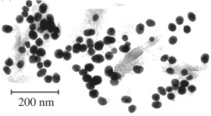
Rudakovskayaらによって合成されたナノ粒子のTEM画像。見てわかるように、ナノ粒子はほぼ球形で、平均サイズは30nmです[76]
コロイド合成
コロイド合成技術は、金属ナノ粒子を合成するためのシンプルで効果的な方法を提供します[82]。コロイド技術は、多くの場合、異なる溶媒を必要とせずに、または室温で実行できるという、ナノ粒子合成の他の技術よりも高いレベルの単純さを提供します[83、84]。合成の基本原理は、水相にさまざまな金属イオンを分散させ、混合物に還元剤を添加し、次に制御された温度で混合して不溶性ナノ粒子を形成することです[39]。コロイド合成経路は、合成に潜在的な有毒溶媒を関与させる必要がないという利点を提供します(ナノ粒子が生物学的使用を目的としている場合に理想的です)。ただし、コロイド経路にはいくつかの制限があります。たとえば、最終的に合成されるナノ粒子のサイズ分布を制御するのが難しい場合があり[85]、ナノ粒子の形状は試薬濃度によって大きく影響を受ける可能性があります[85]。しかし、プラス面としては、ナノ粒子を大量に生産する方が簡単な場合があります[86]。この金属ナノ粒子合成法は長年にわたって使用されており、銀[87]や金[39、88]などのさまざまな種類のナノ粒子の合成に使用されています。
この基本的な方法は、金でコーティングされた酸化鉄ナノ粒子を形成するためのさまざまな合成経路を生成するために進歩および開発されました[83、84、89、90、91、92、93、94、95、96、97]。金でコーティングされた酸化鉄を合成する方法のほとんどは、HAuCl 4 を還元するためにさまざまな還元剤を使用することを中心に展開されます。 酸化鉄の表面に。なだごだ他HAuCl 4 を減らすためにアスコルビン酸を使用する提案された「グリーン」合成ルートを提供します [84] 。 ただし、この方法では、合成に使用されるキャッピング剤(ナノ粒子の外側に結合してナノ粒子のさらなる「成長」を停止する薬剤)がないため、コーティングされたナノ粒子のサイズや形状をほとんどまたはまったく制御できないようです。 [98]。合成されたコーティングされた粒子の形状とサイズに対してより多くの制御を示す方法は、Palらによって提示されています。 [95]この方法では、金塩として酢酸金を使用します。これは、6 nm Fe 3 の表面に還元されます。 O 4 7 nmサイズのAu–Fe 3 を作成するためのナノ粒子 O 4 球形の粒子。 Fe 3 をコーティングするための迅速な方法 O 4 ナノ粒子はRawalらによって提示されています。これには、Fe 3 の分散が含まれます O 4 HAuCl 4 の溶液中のナノ粒子 、次にエタノールと混合する[83]。室温で15分後、反応を停止し、Au-Fe 3 O 4 次に、ナノ粒子を磁石で分離しました。精製された溶液のTEM分析は、生成された粒子のサイズが30〜100 nmの範囲であり、サンプル全体でさまざまな形状を持っていることを示しました。これらの画像は図4で見ることができます。この合成技術はコーティングされたナノ粒子を迅速に生成しましたが、均一な形状とサイズの粒子を生成するための非常に効率的な合成ではないようです[83]。
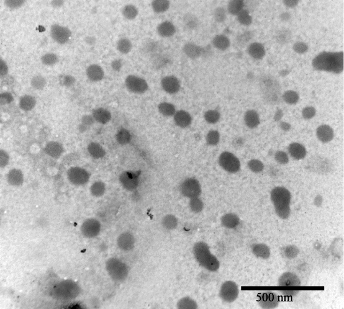
Rawalらによって合成されたナノ粒子のTEM画像。これらのナノ粒子のサイズ分布は20〜100nmです[83]
金塩の還元だけを提供する技術もあれば、ヒドロキシルアミンなどの還元剤を鉄の表面に置くことを好む技術もあります[90、93]。多くの場合、Fe 3 O 4 ナノ粒子は金でコーティングされており、金塩を還元すると標準的な金ナノ粒子も生成されます[74]。したがって、鉄ナノ粒子の表面に還元剤を添加すると、コーティングの効率が向上し、副産物として生成される金ナノ粒子の量[93]。
別の技術は、金を磁性ナノ粒子の表面に播種することを含み、これは、金をナノ粒子の磁気コアの周りに核形成させるより直接的な経路を提供する[91、92、97]。この技術は、溶液中の酸化鉄ナノ粒子よりも小さい金の種子を酸化鉄の表面に結合させることを含みます。 HAuCl 4 の場合 溶液中で還元される、Au + イオンは酸化鉄にシードし、酸化鉄ナノ粒子の周りにシェルを形成します。この金の種まきは、いくつかのグループで成功裏に採用されています。グーンら。ポリエチレンイミンを使用して、Fe 3 の表面への金のシードを制御しました O 4 、完全にコーティングされたナノ粒子を生成します。 [91]ただし、合成されたAu-Fe 3 O 4 粒子は高い多分散性を示し、粒子サイズは40〜110nmの範囲でした。 Levin etal。金のシードに結合するためにオルガノシラン分子で機能化されたコアを使用して、50〜70nmのサイズ範囲の金シェル磁性コアナノ粒子を生成することに成功しました[92]。鉄のコアへの金ナノ粒子の播種は、さまざまなコア形状で実証できます。たとえば、Wang etal。米の形をした「ナノライス」Fe 3 への金の播種を実演 O 4 構造は、金が表面に還元されたときに完全に厚い金の殻になりました[97]。
金属ナノ粒子の生物医学的応用
抗菌剤
細菌感染症は非常に一般的であり、1928年にアレクサンダーフレミングがペニシリンを発見して以来、抗生物質が主要な治療法となっています[99]。ナノメディシンは、将来の治療のために金属ナノ粒子が探求されている、新しい幅広い可能な治療法を私たちに提供します[100]。表1に、抗菌用途で調査されたナノ粒子の一部を示します。その潜在的な用途について検討された1つの材料は銀であり、これはさまざまな生物医学的用途があることが示されています[101]。たとえば、Sreekumar etal。抗菌繊維のネットワークの一部として銀ナノ粒子を利用しました。ナノ粒子のサイズは20〜120 nmで変化し、 Escherichia coli に対する抗菌効果がありました。 銀ナノ粒子を含まない繊維と比較して94.3%もの高さです[102]。アンピシリンなどの抗生物質は、 Eで≤99.9%の殺傷率を達成できることが示されています。コリ [103]、同じ研究はまた、 Eの特定の菌株におけるアンピシリンに対する耐性の出現を報告した。コリ 。これと同じメモで、 E。コリ 銀ナノ粒子に対する耐性を発達させることができます。ただし、この耐性は遺伝的変化ではなく、コロイド状ナノ粒子を凝集させようとする物理的反応です[104]。また、その抗菌特性のために銀を採用している、Holtz等。直径1〜20nmの銀ナノ粒子で「装飾」された60nmのバナジン酸銀ナノワイヤーのシステムを設計しました[105]。このシステムは、3つの黄色ブドウ球菌に対して有望であることが示されました。 菌株であり、興味深いことに、メチシリン耐性黄色ブドウ球菌に対する増殖阻害濃度もはるかに低かった。 (MRSA)抗生物質オキサシリンより。
<図>銀ナノ粒子の合成は、Vermaらによって報告されました。バクテリアに対してナノ粒子を使用した場所: Pseudomonas fluorescens 、 E。コリ 、および真菌カンジダアルビカンス [106]。銀ナノ粒子は、最小発育阻止濃度がそれぞれ4.0μg/ mlと16.0μg/ mlであるアンピシリンやネオマイシンなどの一般的に使用される抗生物質と比較して、3つの株全体で平均最小発育阻止濃度が5.83μg/ mlでした。 Eの菌株に対して。コリ [110]。潜在的に興味深いのは、ナノ粒子が Pに対して表示される特性です。蛍光灯 C。アルビカンス 、どちらも免疫不全患者に病気を引き起こすことに関連しています[111]。さらなる調査により、銀ナノ粒子は、広範な副作用があるアンホテリシンBなどの最も一般的に使用される抗生物質のいくつかよりも病原体を治療するためのより効率的な方法であることがわかるかもしれません[112]。
チオグアニンでキャップされた金ナノ粒子の合成は、Selvarajらによって報告されています。ここで、以下を含むいくつかの細菌に対する強化された抗菌効果: E。コリ 、 Aspergillus fumigatus 、および緑膿菌 [107]。チオグアニンでキャップされた金ナノ粒子は、抗癌剤および抗菌剤として非結合チオグアニンよりも効果的であり、それらの活性は癌治療薬の担体としての潜在的な使用を示していることがわかった。同様に、金ナノ粒子は Corynebacterium pseudotuberculosis に対して抗菌効果があることが報告されています。 [108]、50μg/ mlの用量を使用した平均サイズ25nmのナノ粒子は、20分の曝露後に95%の細菌増殖阻害を示しました。同様に、裸の金ナノ粒子は、 Sを含む様々なグラム陰性菌およびグラム陽性菌に対して抗菌効果を有することが示された。アウレウス 、クレブシエラ肺炎 、および枯草菌 [109]。 1.35μg/ mlのAuNPの用量は、 Sに対して46.4±0.4%、38.3±0.2%、および57.8±0.2%の成長阻害を示した。アウレウス 、 K。肺炎 、および B。枯草菌 それぞれ。
抗ウイルス剤
抗菌用途と同様に、金属ナノ粒子は抗ウイルス用途で有望であることが示されています。表2は、抗ウイルス特性を備えていることが示されているナノ粒子の範囲を示しており、ウイルスの治療に適用できる可能性があります。裸の銀ナノ粒子とコーティングされた銀ナノ粒子の両方[113,114,115,116]は、ナノスケールの範囲にある場合、さまざまな抗ウイルス用途があることが示されています。
<図>B型肝炎(HBV)はウイルス感染症であり、現在世界中で2億5700万人が感染しており、世界保健機関によると2015年に887,000人が死亡しました[121]。小さな(10–50 nm)裸の銀ナノ粒子がHBVの可能な治療法としてテストされ[113]、HBVに効率的に結合し、HBVRNAの生成をさらに阻害することが示されました。作用機序は、AgNPがHBV dsDNA(二本鎖DNA)に結合するためであると仮定されています。 Rogers etal。サル痘ウイルス(MPV)に対する抗ウイルス剤として、裸および多糖類コーティングの両方で銀ナノ粒子の使用を実証しました[114]。ナノ粒子は、12.5〜100μg / mlの濃度範囲でMPVに対してinvitroでテストされました。研究の結果は、使用された多糖類でコーティングされた銀ナノ粒子(Ag-PS-NPs)のすべての濃度が、invitroでMPVによって誘発されたプラーク形成を減らすことができることを示しました。
銀ナノ粒子は、ヒト免疫不全ウイルス(HIV)の治療においても役割を果たす可能性があります[115、116]。 HIVは健康上の大きな懸念事項であり、WHOは2016年の時点で3,670万人がHIVとともに生きていると推定しています[122]。 HIVの治療法を発見し、迅速かつ効率的に実施することが重要です。ララ等。銀ナノ粒子(30〜50 nm)がHIV-1分離株に及ぼす影響を示しており、HIV-1分離株のすべての菌株の阻害を示しています[116]。裸のナノ粒子は全体的なIC 50 を示しました HIV-1に対して0.44mg / ml±0.3であり、ウイルス阻害のメカニズムはウイルス-宿主細胞結合の阻害であることが示されています。具体的には、銀ナノ粒子はgp120タンパク質(外被糖タンパク質)と標的細胞の間の相互作用を阻害します。膜受容体。同じグループによって、ポリビニルピロリドン(PVP)でコーティングされた銀ナノ粒子がHIV-1のヒト頸部組織外植片モデルへのトランスフェクションを防ぐ能力も実証されました[115]。具体的には、0.15 mg / mlのPVPコーティングされた銀ナノ粒子(PVP-AgNPs)は、HIV- IIIB による感染を抑制しました。 およびHIV- AZT-RV 分離します。この濃度のPVP-AgNPは、対照サンプルと比較して、感染部位へのリンパ球(免疫細胞)の増殖も誘発しました[115]。
ウイルスに対して使用されているのは銀とコーティングされた銀ナノ粒子だけではありません。両親媒性硫酸塩リガンドでコーティングされた2nmの金ナノ粒子もHIV-1に対して有効であることが示されました[118]。これらの粒子は、ウイルスの融合プロセスを標的とすることが示され、インビトロでgp120タンパク質に結合し、HIV-1感染を直接中和することが示されました。メルカプトエタンスルホネートでコーティングされた金ナノ粒子(Au-MES)は、おそらく宿主細胞へのウイルスの結合、細胞から細胞へのウイルスの拡散、またはウイルスに対する細胞の感受性の変化を阻害することにより、単純ヘルペスウイルス1型(HSV-1)感染の阻害を示しましたナノ粒子の存在によって誘発される感染症[117]。
ヨウ化銅ナノ粒子(CuI-NP)は、ネコカリシウイルス(FCV)[119]や、さらに興味深いことに豚由来のインフルエンザAウイルス(H1N1)[120]などのいくつかの異なるウイルスに対して抗ウイルス特性を示すことが示されています。 100〜400 nmのCuI-NPは、FCVに対して使用すると抗ウイルス特性を示し、一価のCuイオンが活性酸素種(ROS)の生成に関与し、その後のキャプシドタンパク質の酸化を引き起こしてFCVの不活性化を引き起こすと仮定されました。 H1N1ウイルスは、CuI-NPによっても非常によく似た方法で阻害されることが示されました。つまり、ヒドロキシルラジカルの生成により、タンパク質が分解されます。ただし、これらのラジカルは、感染していない組織に対しても毒性があることが判明する可能性があります。これは、治療の使用が承認される前に決定することが重要です[123]。
イメージング
磁気共鳴画像法(MRI)スキャンは、医療診断に非常に役立つツールであり、明確な解剖学的画像を提供します。 MRIを使用すると、血流、物理化学的特性、および体内の組織や臓器の状態を視覚化できます[124]。造影剤は、診断感度を向上させるためにMRIでよく使用されます[125]。従来使用されていた造影剤はキレートベースですが、現在の造影剤の主な欠点は、細胞に蓄積されたときの生物学的安定性と毒性レベルです[126]。たとえば、一部の造影剤はヨウ素ベースであり、ヨウ素化造影剤への曝露は、その後の甲状腺機能亢進症および顕性甲状腺機能低下症の発症に関連していることが報告されています[127]。造影剤が体に及ぼす可能性のある悪影響を減らすことにより、スキャン効率を改善するための代替案が開発されました[128]。代替案には、MRIスキャン用の造影剤と同様に作用する薬剤と結合している可能性のある金属ナノ粒子が含まれます[129]。図5は、AuNPの治療前と治療後のラット大脳皮質のMRI造影画像です[130]。
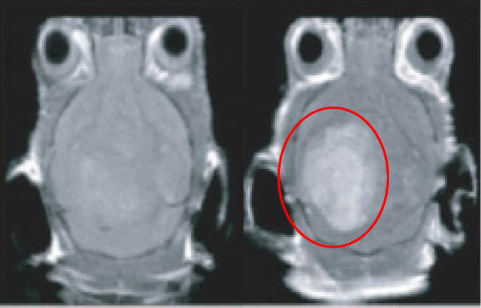
治療前(左)と治療後(右)のラット大脳皮質のMRI造影画像。 AuNPを含む領域は赤で表示されます
表3は、医用画像で使用するために調査されたナノ粒子の一部を示しています。一部のコンピュータ断層撮影(CT)造影剤には、循環半減期の短さ[131]や潜在的な組織損傷[130]などの問題があります。このため、金属ナノ粒子もCTイメージングで使用するために調査されています[132]。 Auナノ粒子は、X線の減衰により、イメージングで有望な用途を示しています[133]。コジマ他は、PEG化デンドリマー(PEG-AuNPs)と結合した金ナノ粒子がinvitroで優れた造影剤となることを示しましたおよび 市販のヨウ素剤イオパミダルと比較したX線コンピュータ断層撮影の場合[134]。 PEG-AuNPは、市販のイオパミダルよりも高いコントラスト効率を示し、体から急速に排泄されました[135]。著者らはまた、PEG-AuNPには光熱療法を可能にする光細胞毒性があることにも言及しました。
<図>Li etal。アテローム性動脈硬化症のイメージングツールとしてコーティングされたAuNPの使用を実証しました。 AuNPは、「単一光子放射型コンピューター断層撮影」(SPECT)と呼ばれるタイプの医用画像に適用されました[136]。このタイプのイメージングは、ガンマカメラの使用と非常に似ていますが、より正確な分析手法を実現するために、スライス、回転、および操作できる真の3D画像を提供できます[136]。修飾されたナノ粒子は、アポトーシスマクロファージを含むアテローム性動脈硬化症のプラークを特異的に標的とし、アテローム性動脈硬化症のプラークを侵襲的に正確に検出するための有用なツールを示しています[136]。
AuNPは、光音響イメージング(PA)の可能性のある薬剤であることが以前に実証されており、高い空間分解能と感度を示しています[137]。 PA relies on the detection of ultrasonic waves which are emitted from tissues when exposed to non-ionizing pulsed laser irradiation [140]. The intensity/magnitude of the ultrasonic emission is responsible for the image contrast, therefore any agent that can both absorb the laser pulses and then give off heat as a result will increase the magnitude of the ultrasonic emission and AuNPs possess the ability to do both of these [141, 142]. AuNPs are potentially better than organic dyes due to the organic dyes’ susceptibility to photo-bleaching and rapid clearing from the blood [143]. AuNPs also have use in cell imaging for examining movement of nanoparticles within cells when conjugated with various cargoes. Figure 6 is a darkfield imaging of A431 lung cancer cells treated with AuNPs that target epidermal growth factor receptor, and the bright areas within the cells are the nanoparticles indicating their locations within the cells [144].

Darkfield imaging of A431 lung cancer cells treated with AuNPs; the bright yellow/orange dots are nanoparticles within the cells
Biomedical Cargo Delivery
Nanoparticles make for an ideal molecule for drug delivery due to the huge surface area to the volume ratio they provide when compared to their bulk material [145]. In addition, it is possible to engineer nanoparticles to either avoid or interact with the immune system in specific ways [146, 147]. For example, it has been demonstrated that an increased hydrophobicity of nanoparticles/sub-groups conjugated to the nanoparticles illicit and increased immune response by measuring cytokine mRNA levels in mice [146]. Focusing in the opposite direction, it has been suggested that nanoparticles can be conjugated with various ligands to directly activate the immune system to target the destruction of a tumor [148] or by accumulation in the liver or spleen for the generation of tolerance or immunity, respectively [147].
Gold nanoparticles have been extensively studied for their delivery of medical cargo, for example, Bhumkar et al. have explored the application of AuNPs for trans-mucosal delivery of insulin. Gold nanoparticles were synthesized in the presence of chitosan, which acts as a polymeric stabilizer [149]. These nanoparticles were then loaded with insulin and administered both nasally and orally to diabetic rats. The results showed an overall reduction in the rat’s blood glucose levels, an indication of successful movement of the nanoparticles through the mucosal membranes and into the bloodstream.
More recently “smart” AuNPs have been employed in PA [138]. These nanoparticles are roughly 10 nm in diameter and are functionalized with citraconic amide moieties which are susceptible to hydrolysis. The citraconic amides are converted into positively charged primary amino acids at a mildly acidic pH, while the surface molecules adopt negative charges at physiological pH [138]. Combined, these two properties cause the “smart” nanoparticles to adopt both positive and negative charges allowing them to aggregate rapidly due to electrostatic attraction. These nanoparticles are referred to as “smart” due to the nanoparticles presenting cancer-specific properties and accumulate rapidly and efficiently in cancer tissues and show a much lower accumulation in normal tissues [150].
Gold nanoparticles can also be used as a delivery system for nucleic acids [153], including oligonucleotides [151] and small interfering RNA (siRNA) [154]. Many different methods have been developed to functionalize AuNPs with nucleic acids, for example, Yonezawa et al. have synthesized gold nanoparticles modified with thiocholine, which then bound to DNA and formed a fusion of wire-like structures throughout the DNA [155]. Sandström et al. demonstrated the ability to bind nucleic acids onto gold nanoparticles [151], and a similar modification has been done by Rosi et al. where tetrathiol-modified antisense oligonucleotides were bound to 13-nm gold nanoparticles [152]. Being able to conjugate nucleic acids to nanoparticles opens up the possibility of targeted gene delivery, which could, for example, lead to genes coding for a specific protein to be delivered to a cell that was either deficient in that protein or could not produce the protein themselves [156]. It has also been exhibited that gold nanoparticles modified with DNA can transfect cancer cells [157] (Table 4).
<図>Anticancer Drug Delivery
Cancer is one of the world’s leading killers with large areas of scientific research being dedicated to the fight against cancer, and nanoparticles offer a new doorway into methods to target and treat cancer. Table 5 presents a selection of nanoparticle/drug conjugates that have been tested for anticancer treatments. Paciotti et al. have investigated the application of PEGylated AuNPs as a carrier for tumor necrosis factor (TNF) which is a cell-signaling protein that possess the ability to induce apoptosis in healthy cells [158]. The Au-PEG-TNF nanoparticles were injected intravenously and agglomerated significantly more in MC-38 colon carcinoma cells compared to other healthy cells/tissues. The TNF not only gave therapeutic action on the MC-38 cells, but also seemed to possess a targeting property, indicated by the lack of agglomeration in healthy cells. Another interesting observation reported was the ability for the Au-PEG-TNF nanoparticles to diminish a tumor mass compared to “free” TNF.
<図>Doxorubicin is a widely used cancer therapeutic agent but has dose-limiting associations with cardiotoxicity. A gold nanoparticle-doxorubicin conjugate has been developed that demonstrates little no to cardiotoxicity to mice while being able to treat cancer [160]. Dixit etal。 demonstrated the selective delivery of folic acid-coated AuNPs into folate receptor (FR) positive cancer cells, whereas when compared with a cell line that did not have folate receptors, uptake was shown to be minimal [159]. These results demonstrated the use of folate to target metal nanoparticles to FR-positive cancer cells for tumor imaging and ablation.
Limitations of Single Metal Nanoparticles and Overcoming Them
The principal obstacle with nanoparticle drug delivery is the ability to direct the nanoparticle to the target area [162, 163]. There are several methods in use for metal nanoparticle targeting such as antibodies [164,165,166] and homing peptides [167, 168]. There are however limitations to these methods, with the biggest being that before they even reach the desired target cells they have to pass through a variety of other barriers, such as blood vessels and the blood-brain barrier [169]. One way to overcome this targeting limitation is to use magnetic nanoparticles [170]. A magnetic nanoparticle-targeting system works by directing the nanoparticles to a target site using an external magnetic field, it has already been demonstrated that the magnetic anisotropy of the nanoparticle is a very important factor for medical treatments [171], with a change in anisotropy being able to the change the efficacy of hypothermia treatments for example [172]. Superparamagnetic metal nanoparticles have this property (they only present magnetic properties while in the presence of a magnetic field) [173]. However, the benefit of magnetic nanoparticles also presents a potential limitation, due to the toxicity of many magnetic materials [31, 174, 175]. Despite iron being approved for various imaging uses [5, 6, 31], it has been suggested in several studies that naked iron oxide nanoparticles may have some adverse effects when used in cell labeling [176,177,178]. One method that can be used to overcome any potential toxicity limitations is to coat the iron core [179]. A range of materials can be used as the coating material:silica [180,181,182], polymers [183, 184], gold [62, 93, 95, 185], or silver [58, 186]. Gold has low pharmaceutical activity [187] and silver has been used in biomedical applications for many years [188, 189],
The combination of a superparamagnetic core with an inert and safe metal coating produces metal nanoparticles with superior characteristics to non-magnetic metal particles [190]. As well as reducing toxicity, the coating also provides the potential for the conjugation of functionalized molecules onto the surface, such as drugs and biomolecules for application in the medical field [97, 140, 152]. It is of note that a core-shell nanoparticle still possesses the properties and uses of a nanoparticle made from the same material as just the shell, but the superparamagnetic core gives the ability to direct the nanoparticle in the body [191]. For example, a gold nanoparticle with an antibody is classified as a targeting nanoparticle, introducing the core would classify the nanoparticle as a directed targeting nanoparticle [173].
Current Medicinal Uses of Gold-Coated Iron Oxide Nanoparticles
Core-shell superparamagnetic nanoparticles have already been assessed for their biomedical uses, with a wide range of uses already being applied [192] and with a majority of research investigating into the use of gold as a shell for the nanoparticles, in part due to its biocompatibility and ability to easily bind to a variety of materials. As such, this section will deal exclusively with gold shell nanoparticles. One of these uses is as a magnetic carrier for drug targeting [192,193,194,195,196]. Kayal and Ramanujan have tested an in vitro apparatus that simulates the human circulatory system as a test for the magnetic delivery of gold-coated iron oxide nanoparticles (Au-Fe3 O 4 ) loaded with doxorubicin [194]. Their system had various magnetic fields of increasing strength next to a capillary through which the doxorubicin-loaded particles were passed. A significant percentage of these nanoparticles were captured within the magnetic fields, strongly indicating the potential for the use of magnetic nanoparticles in drug delivery. Another use for a targeted system is the application of Au-Fe3 O 4 nanoparticles in photothermal therapy. Bhana et al. demonstrated the use of a core-shell system used in combination therapy deployed against two different cancer cell lines; head and neck (KB-3-1) and breast (SK-BR-3) with a reported decrease in cell viability of 64% when they exposed cell lines to a combined photothermal and photodynamic therapy, compared to each modality used on its own [197]. In photothermal therapy, gold nanoparticles are coated with a ligand, such as PEG [142], and these nanoparticles are irradiated with a laser, with a wavelength that matches the UV-vis λ -max of the gold nanoparticles [194]. The nanoparticles vibrate at the laser’s frequency which causes heat to be released causing the death of the surrounding tissue [198], introducing a core which is superparamagnetic can allow for a more accurate targeting for use in this therapy. Similarly, it has been reported by Kirui et al. that gold hybrid nanoparticles were deployed against SW1222 colorectal cancer in photothermal therapy, showing an increased case of cellular apoptosis after therapy, with their conclusion being that the cells showed an increased uptake, leading to a reduced laser power required to reach threshold therapeutic levels [199]. The use of core-shell nanoparticles for photothermal therapy of cancer has also been reported by other groups [200, 201].
Metal nanoparticles have already shown to have a place in contrast imaging, for example core-shell nanoparticles can also be used in T1- and T2 -weighted imaging in MRI [202]. Research by Cho et al. demonstrated that gold-coated iron nanoparticles can be successfully used in MRI imaging, as well as opening the route for conjugating various ligands for use in biosensors [202]. A magnetic carrier capable of imaging and photothermal therapy has been reported by Cheng et al. They demonstrated the magnetic targeting of multi-functional nanoparticles to a tumor in a mouse model, which could be imaged inside the tumor and showed a reduction in the tumor size when combined with photothermal therapy [203]. It is also of note that in this work, both the nanoparticle dosage (1.6 mg/kg) and laser power (1 W/cm 2 ) are among the lowest applied for in vivo photothermal therapy. Moreover, there was no obvious toxicity from the nanoparticles reported. Table 6 presents some of the currently reported uses of core-shell nanoparticles.
<図>Another medical area where such core-shell metal nanoparticles have been suggested to make an impact is in directed enzyme prodrug therapy (DEPT) [170, 191]. DEPT is a promising method of cancer treatment, with several therapies making it through to clinical trials [207, 208]. The main principal of DEPT is the targeted delivery of a prodrug-activating enzyme to a tumor site. Upon arrival at the tumor site, the enzyme enters the target cells where it can later activate an administered prodrug. However, the efficacy of the therapy depends on the ability to direct the enzyme to the tumor site, with current directional techniques relying on passive targeting methods such as viruses [207, 209] or antibodies [210, 211], rather than an active targeting system for enzyme delivery. A novel therapy proposed by Gwenin et al. potentially overcomes the targeting issue [170, 212]. This approach involves conjugating a genetically modified prodrug-activating enzyme onto the surface of a gold-coated iron oxide superparamagnetic nanoparticle (AuMNP), then directing the AuMNP-enzyme conjugate to the target site using a magnetic field to increase the efficacy of the targeted therapy. Figure 7 presents some of the uses of a core-shell nanoparticle.

A pictorial representation of the applications of core/shell nanoparticles
結論
In brief, single metal nanoparticles have been shown to currently possess a wide range of biomedical applications, with more application for these nanoparticles being discovered. One of the limiting factors that these nanoparticles face in medical treatments is to find a way for precise accurate targeting of areas within the body, be it for targeting of a drug delivery or for therapies involving the nanoparticles directly. A way to overcome this is to employ a magnetic core to create core-shell nanoparticles that can then be directed around a body using a magnetic field. There are a variety of methods that can be used to synthesize these core-shell nanoparticles, with each method having its own advantages and disadvantages. There remain many obstacles for core-shell nanoparticles before they can be routinely applied in the medical field and these include
- 1)
Achieving a synthesis route which produces easily repeatable results;
- 2)
Producing particles of a set size [22,23,24] and shape [25,26,27,28]; and
- 3)
Producing large enough quantities to make it economically viable [29].
Another key factor is the relatively unknown toxicity of some nanoparticles over an extended period of time due to how relatively new the field of research is.
データと資料の可用性
Not applicable
略語
- (o/w):
-
Oil-in-water
- (w/o):
-
Water-in-oil
- (w/sc-CO2 ):
-
Water-in-supercritical CO2
- AgNP:
-
Silver nanoparticle
- Ag-PS-NPs:
-
Polysaccharide-coated silver nanoparticles
- Au-Fe3 O 4 :
-
Gold-coated iron oxide nanoparticle
- Au-MES:
-
Mercaptoethane sulfate-coated gold nanoparticle
- AuNP:
-
Gold nanoparticle
- Au-PEG-TNF:
-
Polyethylene glycol-coated tumor necrosis factor-loaded gold nanoparticles
- CT:
-
Computed tomography
- CTAB:
-
Cetyltrimethylammonium bromide
- CuI NPs:
-
Copper-iodine nanoparticles
- DNA:
-
Deoxyribonucleic acid
- FCV:
-
Feline calicivirus
- FR:
-
Folate receptor
- Gp120:
-
Glycoprotein 120
- H1N1:
-
Influenza A of swine origin
- HBV:
-
Hepatitis B virus
- HIV:
-
Human immune-deficiency virus-1
- HSV-1:
-
Herpes simplex virus 1
- KB-3-1:
-
Head and neck cancer
- MC-38:
-
Colon carcinoma
- MPV:
-
Monkeypox virus
- MRI:
-
磁気共鳴画像法
- PA:
-
Photoacoustic imaging
- PEG:
-
ポリエチレングリコール
- PVP-AgNP:
-
Polyvinylpyrrolidone-coated silver nanoparticle
- RNA:
-
Ribonucleic acid
- ROS:
-
活性酸素種
- siRNA:
-
Small interfering ribonucleic acid
- Sk-BR-3:
-
Breast cancer
- SPECT:
-
Single-photon emission computed tomography
- TNF:
-
Tumor necrosis factor
ナノマテリアル
- ドラッグデリバリーを強化するためのナノファイバーとフィラメント
- 改善された診断および治療用途のための多機能金ナノ粒子:レビュー
- 癌治療のためのナノ粒子:現在の進歩と課題
- 生体適合性FePO4ナノ粒子:ドラッグデリバリー、RNA安定化、および機能的活性
- 合成および生物医学的応用のための蛍光ナノ材料の進歩と挑戦
- コバルトをドープしたFeMn2O4スピネルナノ粒子の調製と磁気特性
- 癌用途のための細胞ベースのドラッグデリバリー
- 黒色腫の標的化学療法治療のための薬物送達担体としての131I追跡PLGA-脂質ナノ粒子
- ナノテクノロジー:invivoイメージングシステムから制御されたドラッグデリバリーまで
- In VivoCTイメージングおよび腎クリアランス特性のための新しい生体適合性AuNanostars @PEGナノ粒子
- 3Dプリントされたマイクロロボットはドラッグデリバリーの約束を保持します