


ナノテクニックは癌幹細胞を不活性化する
要約
現在の腫瘍学の課題の1つは、癌幹細胞の同定とそれらの特異的阻害が可能な治療手段の探索です。この論文は、エーリッヒ癌細胞の表現型の特徴に関するデータを、腫瘍増殖の便利で追跡しやすいモデルとして提示しています。エーリッヒ癌の一部としての癌幹細胞の証拠とCD44 + の重要性 およびCD44 – このタイプの腫瘍の成長を維持する亜集団が実証されました。エーリッヒ癌CD44の高い(10倍)腫瘍形成活性 + CD44と比較した場合の細胞 – 細胞が証明されました。このペアの比較では、CD44 + 細胞は、CD44 high の腹腔内で生成する可能性が高かった 、CD44 + CD24 – 、CD44 + CD24 + CD44 + のプール内の癌幹細胞の存在を強調する細胞亜集団 セル。
この研究では、希土類オルトバナジン酸塩GdYVO 4 のナノ粒子を含む合成ハイブリッドナノ複合体の能力 :Eu 3+ 腫瘍の成長を抑制し、腫瘍のある動物の生存率を高めるためのコレステロールが確立されました。腫瘍抑制効果への特別な貢献は、その各コンポーネントによって行われます。エーリッヒ癌細胞を2成分ハイブリッド複合体で処理すると、最も腫瘍形成性の高いCD44 high の濃度が最大に低下しました。 CD117 + の数が同時に増加する細胞 対照と比較した場合、腫瘍増殖の強度を74.70±4.38%減少させた細胞。
背景
悪性腫瘍の成長の問題は、依然として医学において最も緊急の問題の1つです。ここ数十年で、癌の新しい治療法の開発にいくらかの進歩がありました。これは、癌の古典的な概念の改訂と、無制限の自己複製が可能であり、多くの表現型マーカーによって識別できる癌幹細胞(CSC)の発見によるものです。これらの細胞のほとんどは、放射線療法および化学療法に耐性があり、悪性腫瘍の増殖と転移の再発を引き起こします。抗がん療法の新しい方法が習得されています。つまり、正常組織への損傷を最小限に抑えて腫瘍細胞を選択的に不活化することができます[1]。
CSCは、1997年にM.Dickチームによって初めて識別および記述されました[2]。著者らは、免疫不全NOD / SCID(非肥満性糖尿病-重症複合免疫不全症)マウスに移植した場合、細胞の総集団の0.01〜1%である亜集団が白血病を引き起こす可能性がある急性骨髄性白血病を調査しました。これらの腫瘍誘導細胞は、表現型的にCD34 + として特徴づけられました。 CD38 – 。 2003年、M。Al-HajjとM.S. Wichaは、固形のヒト乳がん(BC)のCSCを特定することに成功しました[3]。原発性乳がんの分離されていない集団は、5×10 4 の濃度でNOD / SCIDマウスに投与された場合、100%の症例(10/10)で腫瘍形成能を示すことがわかっています。 細胞/マウス。投与される細胞の濃度を1×10 4 に下げる 細胞/マウスは腫瘍形成活性を4倍低下させました(3/12)[3]。 CD24 + CD44 + さまざまな用量(2×10 4 )で投与した場合の割合 100細胞/マウスまで)は腫瘍の成長を許しませんでした。これにより、CD44 + CD24 – /低亜集団は有意に高い腫瘍形成活性を有し、10 3 を投与した場合の100%の症例で腫瘍の形成を示しています。 細胞/マウス。腫瘍を形成する最も顕著な能力は、CD44 + の亜集団に固有のものでした。 CD24 – / lo ESA + 表現型。これらの細胞のうち200個だけをマウスに投与すると、注射後5か月で100%(4/4)で固形腫瘍が形成されました[3]。これらの研究は、乳がん生検サンプルの特定の集団が無血清培養でinvitroでマンモスフィアを形成する能力を示したPontiD。etal。によって継続されました[4]。得られたマンモスフィアの細胞のほとんどはCD44 + でした。 / CD24 – / low SCID(重症複合免疫不全症)マウスに投与した場合の表現型およびinvivoでの腫瘍形成能の増加。この亜集団で腫瘍を形成する能力は、伝統的に移植された乳がんMCF7の系統と比較して1000倍高かった[4]。ただし、著者は、CD44 + の20%のみを示しています。 CD24 – / low 細胞には自己複製能力がありました。これは、この亜集団の不均一性、すなわち、細胞の機能を決定する追加のマーカー(ESA、ALDH)の存在に起因する可能性があり、CD44発現速度にも関連している可能性があります。過去数年間に発表された論文は、マーカーの発現が高い(CD44 high )CSCを示しています。 )最も高い腫瘍形成活性を持っています[5、6]。 5×10 5 の同所移植において CD44 高 -RAS変換およびCD44 低 NOD / SCIDマウスに対する細胞では、亜集団CD44が 低い であることがわかっています。 腫瘍形成能は低く(腫瘍は症例の30%で形成された)、CD44は 高い 細胞は100%の症例で腫瘍を形成することができました[6]。
公開されたデータを要約すると、乳がん細胞の亜集団の分化行は次のように表すことができます。
$$ \ mathrm {C} \ mathrm {D} {44} ^ {\ mathrm {high}} \ to \ mathrm {C} \ mathrm {D} {44} ^ {+} \ mathrm {C} \ mathrm { D} {24} ^ {\ hbox {-}} \ to \ kern0.5em \ mathrm {C} \ mathrm {D} {44} ^ {+} \ mathrm {C} \ mathrm {D} {24} ^ {+} \ to \ mathrm {C} \ mathrm {D} {44} ^ {\ hbox {-}} \ mathrm {C} \ mathrm {D} {24} ^ {+} $$他のマーカー、特にSca-1 + を持つ多数の細胞 CSCの段階を主張します。 Sca-1ノックアウトマウスにおける腫瘍増殖の減少に関するデータは、Sca-1 + の腫瘍開始の役割の仮説を支持していることを示しています。 腫瘍形成の初期段階にある細胞[7]。最近、研究者のより多くの注目が、CSCだけでなく、それらの付属調節微小環境を作る細胞によっても引き付けられています。 CD117 + 血液幹細胞のプールで伝統的に検出される細胞は、それらの中で特に注目に値します[8]。ヒト乳がん細胞の総集団は、CD117 + を伴ういわゆる間質関連線維芽細胞で構成されています。 表現型。それらは腫瘍の成長をサポートし、その血管新生を促進します[9、10]。幹細胞のエーリッヒ癌(EC)集団における存在の仮定、CD44 + の腫瘍形成能の研究 CD117 + の割合と役割 腫瘍の発生を維持する細胞には、追加の証拠が必要です。
インビボでCSCを継代するための実験のほとんどは、SCIDまたはNOD / SCIDマウスで実施された。これらのマウスは、ヒト細胞の異種移植に対する免疫反応に反応しません。さまざまな治療薬の抗腫瘍活性を研究および評価するための適切で関連性のある実験モデルの探索が進行中です。それらの1つは、マウスの自然発生乳がんで得られたECのinvivo移植腫瘍細胞株です[11]。ただし、EC細胞の亜集団組成とその表現型の特徴、CSCの存在、およびこのタイプの腫瘍の成長を維持する上での重要性については、事実上出版物はありません。 ECとBCの組織形成的類似性を考慮すると、シミュレートされた腫瘍の開始と発生には、癌細胞の増殖を制御する同じ遺伝子と、腫瘍マーカータンパク質の発現につながる同様の生化学的経路が関与していると考えられます。ただし、EC集団におけるCSCの存在に関する仮定と、それらの腫瘍形成能の研究には、本研究の目的の1つである追加の証拠が必要です。
現在の腫瘍学の緊急の問題は、CSCを特異的に認識するだけでなく、不活性化する薬剤を見つけることです。問題を理解するという概念そのものが、「セラノスティックス」(治療+診断)の瞬間の方向に形成された基礎でした[12]。セラノスティクスの枠内で、癌の診断と治療を同時に行うための医薬品とツールを使用する技術的アプローチが開発されています。セラノスティクスの方向性の1つは、金ナノ粒子を腫瘍部位にターゲティングし、光熱療法を行うことです[13]。腫瘍細胞を同定するための他のアプローチは、量子ドットの使用であり、これは、インビボでの腫瘍のモニタリングを可能にする強力な光学的造影剤である[14]。インビボでヒト胚性幹細胞を非侵襲的に視覚化する量子ドットの能力は、それらの可能な生物医学的応用を支持することを証明している[15]。
最近、希土類元素、すなわち希土類金属(特にバナジウムとその化合物)のナノ粒子(NP)で活性化された誘電体材料とワイドゾーン半導体に基づくナノ発光団に大きな注目が集まっています[16]。これらの材料は、高い光安定性、発光の大きなストークスシフト、シンチレーション効果の欠如、および特徴的な狭い発光バンドの安定性を備えています。これにより、バナジウム化合物の抗腫瘍効果が知られている。したがって、二塩化バナジウムは、核ヘテロクロマチンへの蓄積とそれに続く有糸分裂の有糸分裂異常の一過性抑制の誘導の結果として細胞増殖を有意に阻害し、 S 後期に細胞を蓄積させることが示されています。 および G 2 フェーズ[17]。悪性腫瘍の治療に有望なのは、オルトバナジン酸塩GdYVO 4 の希土類ベースのNPに基づくハイブリッドナノ複合体の使用です。 :Eu 3+ ウクライナ国立科学アカデミーのシンチレーション材料研究所で開発されたコレステロール[18]。
それらの作成の目的は、標的細胞膜に親和性を有するナノ複合体の組成物中に存在するために、抗癌剤の治療効果を高めることであった。 1つはコレステロールで、これは癌細胞を増殖させて生体膜を構築することにより、血流から積極的に「引き抜かれ」ます。これは、遊離血流コレステロールと結合できる多数の腫瘍細胞SR-B1(スカベンジャー受容体、クラスBタイプI)およびカベオリン-1(Cav-1)受容体の表面に存在することによって促進されます[19]。 。
したがって、この研究の目的は、CSCの兆候を示すものを含む、EC細胞の亜集団組成、およびハイブリッドナノ複合体による前処理後の腫瘍形成活性を特定することでした。
メソッド
実験は生後8ヶ月の雌のBalb / Cマウスで行われました。マウスは、動物園の標準状態(室温20±2°C、相対湿度50〜70%、明暗サイクル12:12 h)で飼育されました。すべての実験プロトコルは、ウクライナ国立科学アカデミー、ハリコフ、ウクライナの低温生物学および低温医学の問題研究所の動物倫理委員会によって承認され(2017年1月23日のrec。no。1)、実験動物の使用(Strasbourg、1986)、第1回ウクライナ国立生物倫理会議(Kiev 2004)によって承認されました。
EC細胞のinvivoでの培養
エーリッヒ癌(EC)細胞は、Balb / Cマウスの腹腔(PC)で継代されました。腹水EC細胞で凍結保存されたものを初代培養として使用しました[20]。解凍後、EC細胞をin vivoで3回再移植し、凍結解凍の要因の影響を軽減し、ネイティブ細胞の形態学的および機能的特徴を取得しました[21]。 「安定化」されたEC細胞は、3×10 6 の用量で腹腔内注射されました。 0.3 mlの生理食塩水中の細胞/マウスで、invivoで7日間培養しました。 7日後、軽いエーテル麻酔下で実験動物を実験から除外しました。 PCからの腹水は、内径2.69 mmの針から注射器で採取され、10mlの測定チューブに入れられました。絶対細胞数は、ゴリヤエフチャンバーでカウントされたEC細胞の数で腹腔腹水(ml)に蓄積された量を拡大することによって決定されました。総数が35.00×10 7 まで増加 7日目までのマウスのPCにおけるEC細胞は癌腫発生の基準でした[21]。将来的には、まさに細胞が研究の対象となりました。
ECサブポピュレーションの表現型評価
CD44(FITC)(no。553133、クローンIM7)、CD117(FITC)(no。 553354、クローン2B8)およびSca-1(FITC)no。 553333、クローンE13-161.7)、およびCD24(PE)no。 553262、クローンM1 / 69)を製造元の指示に従ってください。対照として、同じアイソタイプの非免疫FITCおよびPE標識モノクローナル抗体(「BDBiosciences」)を添加したサンプル、いいえ。 553988、クローンA95-1およびno。試験したマーカーに対する抗体として、553989、クローンA95-1)を使用した。免疫表現型の二重染色は、CD44(FITC)およびCD24(PE)モノクローナル抗体を使用して実行されました。 CD44マーカーの平均蛍光が10 3 高い細胞 (対数目盛による)CD44 high サブポピュレーション。結果の記録と分析は、「WinMDi 2.9」ソフトウェア(Joseph Trotter、ラホーヤ、米国)を使用して実行されました。
CD44の分離 + 免疫磁気ソーティングを使用したEC細胞の割合
CD44マーカーの発現レベルが高いCSC(CD44 high )、CD44 + の異種集団で構成されています 細胞は、最高の腫瘍形成活性を有し、磁気ソーター(BDTM Imagnet)を使用して全EC集団から分離されました。 CD44 + を分離するには 派閥では、製造元のプロトコルに従って、マーカーCD44(BD、558739)および二次マウスIgG1磁性粒子-DM(BD、557983)に対して一次非標識モノクローナル抗体が使用されました。 CD44 + の分離純度 総EC人口からの細胞は90%でした。
全集団の細胞および単離されたCD44の細胞の腫瘍形成活性の測定 + およびCD44 – -ECフラクション
全集団および単離されたCD44 + の腫瘍形成能 およびCD44 – EC画分は、上記のinvivoでの培養方法によって比較分析されました。実験のセットアップを図1に示します。
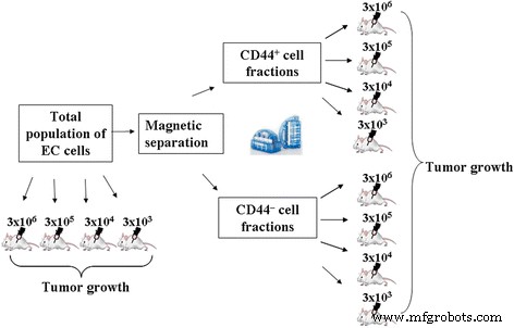
全集団と単離されたCD44 + の腫瘍形成能を比較分析する際の実験計画 およびCD44 – ECフラクション
最初の一連の実験では、総集団と単離されたCD44 + の腫瘍形成能を評価しました。 およびCD44 – EC開始に使用される標準用量(3×10 6 )で動物に投与する場合のEC画分 0.3 mlの生理食塩水中の細胞)。
動物は以下のグループに分けられた( n =10):
-
グループ1.1– EC細胞の総集団の投与(3×10 6 細胞/動物)
-
グループ2.1– CD44 + の管理 EC細胞の割合(3×10 6 細胞/動物)
-
グループ3.1–CD44の管理 – フラクションEC細胞(3×10 6 細胞/動物)
各実験グループに接種してから7日後に、動物のPC内の細胞の総数をカウントし、細胞の表現型の特徴を評価し(上記のとおり)、CD44 high / CD117 + CD44 high の比率として決定されたEC細胞の比率 CD117に対するパーセンテージ + セル[22]。総EC集団および単離されたCD44 + の細胞の増殖能 およびCD44 – 画分は、次のデータに基づいて推定されました:培養時間中の細胞集団余剰の多重度係数(MF)、 M =N / N 0 ;および時間倍増(TD)、TD =(log 2 2)* t / [log 2 ( N / N 0 )]、ここで t は細胞培養の時間(h)、 N t でのセルの数です 時間; N 0 は初期セル番号[23]です。
実験の2番目のセットでは、総集団および単離されたCD44 + の投与された細胞の推定最小用量がありました。 およびCD44 – EC画分、腫瘍増殖を誘発します。全細胞懸濁液および単離されたCD44 + およびCD44 – EC画分を3×10 6 の用量でマウスに腹腔内投与しました。 、3×10 5 、3×10 4 、および3×10 3 0.3 mlの生理食塩水中でマウスあたりの細胞数を測定し、PCで7日間培養しました。
この一連の実験で使用された動物は、次のグループに分けられています( n =10):
-
グループ1.1– EC細胞の総集団の投与(3×10 6 細胞動物)
-
グループ1.2– EC細胞の総集団の投与(3×10 5 細胞/動物)
-
グループ1.3– EC細胞の総集団の投与(3×10 4 細胞/動物)
-
グループ1.4– EC細胞の総集団の投与(3×10 3 細胞/動物)
-
グループ2.1– CD44 + の管理 EC細胞の割合(3×10 6 細胞/動物)
-
グループ2.2– CD44 + の管理 EC細胞の割合(3×10 5 細胞/動物)
-
グループ2.3– CD44 + の管理 EC細胞の割合(3×10 4 細胞/動物)
-
グループ2.4– CD44 + の管理 EC細胞の割合(3×10 3 細胞/動物)
-
グループ3.1–CD44の管理 – フラクションEC細胞(3×10 6 細胞/動物)
-
グループ3.2–CD44の管理 – フラクションEC細胞(3×10 5 細胞/動物)
-
グループ3.3–CD44の管理 – フラクションEC細胞(3×10 4 細胞/動物)
-
グループ3.4–CD44の管理 – フラクションEC細胞(3×10 3 細胞/動物)
すべての実験グループで、PC内の細胞の総数と腹水を発症した動物の総数をEC接種の7日後に測定しました。
ナノ複合体の合成
1.30 g / lの濃度の球状ナノ粒子(NP)(直径2〜3 nm)と0.55 g / lの濃度のヒツジコレステロール(「Acrosorganics」、ベルギー)を含むハイブリッドナノ複合体は、シンチレーション材料研究所で合成されました。報告されているようにウクライナ国立科学アカデミー(ハリコフ)の[18]。希土類元素のオルトバナジン酸塩に基づくNPGdYVO 4 :Eu 3+ 1.30 g / lの濃度の球形のを[24]に記載されているように調製しました。オルトバナジン酸塩をベースにしたコロイド水溶液は、膜「CelluSepH1」3.5KDaを使用した透析によって不純物が精製されています。
ハイブリッドナノコンプレックスでは、負に帯電したNPは、ファンデルワールス力と疎水性相互作用により、コレステロール粒子の周辺に沿って局在します。 NPは、静電相互作用を介してナノ複合体を安定化します。合成されたナノ複合体のサイズは100nmを超えません。さらに、NPは抗酸化特性を示し、酸化を受けません。この事実は、活性酸素種に関連してコレステロールの水性分散液の耐性の上昇に寄与する。ハイブリッドナノコンプレックスの概略構造を図2に示します。
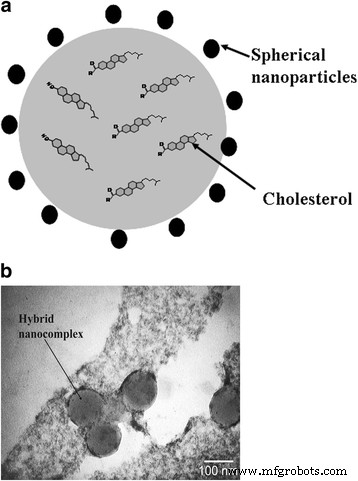
ハイブリッドナノコンプレックス: a 概略図と b カーボンネットワーク上に配置されたコレステロール水溶液から調達されたハイブリッドナノ複合体の透過型電子顕微鏡顕微鏡写真
インビトロ研究中に細胞内のハイブリッドナノ複合体の蓄積を記録するために、疎水性蛍光色素1,1'-ジオクタデシル-3,3,3 '、3'-テトラメチルインドカルボシアニン過塩素酸塩(DiI)をコレステロール水性分散液にさらに導入することができます。局所発光分光法で、複合体の細胞膜への統合のダイナミクスを、モノマー-「J-凝集体」発光バンドの比率によって評価できるようにします[25]。私たちの以前の研究では、ハイブリッドナノコンプレックスは、EC集団全体の細胞の10%以下に、そして事実上、単離されたCD44 + のすべての細胞に統合できることが示されています。 発がん性が最も高い画分。これにより、癌細胞におけるナノ複合体の局所的な蓄積を特定する方法として、この修飾(NP +コレステロール+ DiI)でハイブリッドナノ複合体を使用できるようになります[26、27]。
ナノマテリアルによるEC細胞の前処理
ハイブリッドナノコンプレックスまたはNPを含むEC細胞の全懸濁液を、5%グルコースの溶液(「注入」CJSC、キエフ)で、室温で3時間インキュベートしました。このようなインキュベーション時間は、ナノ複合体を細胞に結合するための最適なものとして以前に発見されました[26]。
ナノマテリアルで前処理されたEC細胞の以下の変異体がテストされました:
-
オプション1–900μlのEC細胞(1×10 7 )100μlの球状NP(1.3 g / l)が追加されました。
-
オプション2–900μlのEC細胞(1×10 7 )100μlのハイブリッド複合体(球状NP(1.3 g / l)+コレステロール(0.55 g / l))を追加しました。
対照は、EC総集団の細胞であり、ナノコンポジットで処理せずに5%グルコース溶液中でインキュベートした。各実験群の動物数は20頭以上でした。
インキュベーション後、すべてのテストグループのEC細胞を、遠心分離(300 gで10分)により生理食塩水(1:1)で3回洗浄しました。
ナノマテリアルによる前処理後のEC発生の強度は、3×10 6 の用量で腹腔内注射することによって評価されました。 0.3mlの生理食塩水中の細胞。すべての研究グループでEC細胞接種後7日で、次のことが決定されました。
-
腹腔内のEC細胞の総数(TN)。
-
式Ri =(TN(c)– TN(e))によるEC成長の阻害率(Ri):TN(c)×100%、ここでTN(c)-PC内のEAC細胞の総数対照群、テネシー州(e)-実験群のPC内のEAC細胞の総数。
-
ECの成長率(Rg)は、式Rg(e)=Rg(c)– Riを使用して計算されました。ここで、Rg(e)-実験動物グループの腫瘍の成長率。 Rg(c)-対照群の腫瘍の成長率、Ri-実験群の動物におけるEC成長の阻害率。対照におけるEC成長の阻害率を100%とすると、EC成長の阻害はありませんでした。
-
CD44 高 / CD117 + 比率(CD44 高 の比率 CD117に対するパーセンテージ + セル)。
-
動物の生存は、未処理およびすべてのタイプのナノコンポジットEC細胞で処理された腹腔内注射後20日目まで評価されました。
統計的処理は、ノンパラメトリックなマンホイットニー U を使用して実行されました。 Statistica6.0ソフトウェアでテストします。 P では、差異は統計的に有意であると見なされました。 <0.05。
結果
得られた結果は、CD44、CD24、Sca-1マーカー、および微小環境の付属調節要素(CD117)に起因する可能性のあるものを表面に持つEC細胞の不均一な集団の存在を示しています。総ECプール(グループ1.1)におけるこれらの特性を持つ細胞の濃度は、表1に示され、EC亜集団の組成に関する以前の調査結果と完全に一致しています[28]。事実上すべてのEC細胞でSca-1構造を同定することにより、この腫瘍タイプの用途の広いマーカーと見なすことができます。
<図>CSCの表現型の同定に関して最も有益なのは、CD44分子の発現であり、これ自体または他の表面マーカーと組み合わせて、ECを含むさまざまな腫瘍からこの細胞集団を分離するために使用されます。古典的な概念によると、乳がんの発生中の腫瘍細胞の分化は、CD24マーカーを発現する細胞の段階的な消失と出現を伴うCD-44受容体の発現低下を伴います[3]。
EC中のCSCの役割の候補は、CD44 high の細胞である可能性があります。 CD44 + の一部である表現型 CD24 – - 人口。 CD44 + の亜集団の機能的活動に対するECの依存性に関するこの仮定 CD44 + によって誘発された腫瘍増殖の強度を評価するときに、細胞をテストしました。 およびCD44 – 派閥とECの総人口。表1は、最高の腫瘍誘発活性がCD44 + の細胞に固有のものであることを示しています。 分数。実際、3x10 6 を投与した後 CD44 + 細胞(グループ2.1)、PCの細胞の絶対数は、EC細胞の総集団(グループ1.1)の23倍、CD44 – の場合の105倍でした。 画分が投与された(グループ3.1)。
これにより、発生中の腫瘍の量的組成だけでなく質的組成の変化も見られた。 CD44 + の割合 CD44 + を主成分とする腹水を形成した セル、つまりCD44 高 、CD44 + CD24 – 、およびCD44 + CD24 + 細胞。その上、CD44の濃度は 高い セルは、グループ1.1と比較して2倍高く、グループ3.1では16倍高かった。 CD44の割合 – 対照的に、より成熟した細胞、すなわちCD44 – を持つ細胞を含む腫瘍を形成しました CD24 + 表現型。グループ3.1の細胞の亜集団組成の非常に再分布により、PC内の細胞の最小絶対含有量が明らかに決定されました。
重要なのは、CD117 + を持つ亜集団のEC細胞間に存在するという確立された事実です。 マーカー。 CD117の分子は膜貫通型チロシンキナーゼ受容体です。通常の条件下では、対応するリガンド、すなわち幹細胞成長因子(SCGF)によって活性化されます[29]。腫瘍病理学では、c-KIT受容体のリガンドオン依存性活性化が起こります。これは、ほとんどの場合(症例の最大92%)、c-kit腫瘍遺伝子変異の結果であるか、この受容体機能の調節の無秩序なメカニズムによって引き起こされます。 [30]。
CD117を検討する + 腫瘍微小環境の細胞としての細胞は、CD117 + の有無に対する腫瘍増殖強度の依存性の確立された事実 細胞とCD44 high との濃度の関係 セルは論理です。表1に示すように、PCに総細胞集団(グループ1.1。)を導入してECを開始すると、34.80±1.27×10 7 が形成されました。 CD44 高 のセル / CD117 + 比率。0.02相対単位に相当します。
CD44の腫瘍形成能 – 割合は4分の1であり(グループ3.1)、CD44 high の減少によって明らかになりました。 / CD117 + グループ1.1と比較した場合、同程度(4倍)の比率。 CD44のこの変更 高 / CD117 + 指数は主にCD44 high の減少によるものでした CD117 + の含有量が減少した背景の濃度(8.5倍) セル(2回)も。
CD44 + によって生成される腹水の成長の強度を評価する場合 画分では、PC内の細胞の総数の有意な増加(グループ1.1と比較した場合はほぼ24倍)が認められました。同様に、重要なのは、CD44 high の2倍の超過です。 CD117の集中と欠如 + 細胞。 CD44の初期資料 + データのフローサイトメトリー分析による画分(培養前)、CD44の含有量 高 細胞はEC細胞の総集団よりも15倍高かった(データは提示されていない)。
上記を要約すると、ECの開始における重要な役割は、CD44マーカーの発現率が高い(CD44 high )CSCによって果たされていると主張することができます。 )、CD117 + の最も重要な機能の1つです。 亜集団は、CD44 high の腫瘍形成活性の調節(「抑制」)です。 細胞。 CD117 + がない 細胞(グループ2.1)は、CD44 + のプール全体の増殖および分化の可能性を倍増させるようです。 セル、PC内のセルの総数の大幅な増加を引き起こします。
EC細胞とCD44 + の総プールの増殖能の分析 派閥はこの解釈を支持します。グループ2.1のPCで培養された全集団の増殖係数(MF)が示されています。グループ1.1と比較した場合、7日間でほぼ24倍に増加しました。これは、CD44 + から成長した腹水細胞の集団を特徴づける可能性のある、グループ1.1の24.47±2.75時間からグループ2.1の14.70±1.35への細胞時間の倍増の減少を伴いました。 より活発に増殖しているものとしての割合(表1)。
CD44 + の特別な役割を証明するため 最小用量でもECを投与した場合の腫瘍の開始と維持における細胞、単離されたCD44 + の腫瘍形成能を比較評価することは興味深いことでした。 およびCD44 – さまざまな濃度で投与された場合の画分。 3×10 6 の導入後、 総EC集団の細胞では、100%の動物で腫瘍の成長が観察されました(10/10)(表2)。投与される細胞の用量を10倍減らす(3×10 5 ) resulted in a proportional decrease in absolute number of cells in the PC, tumor developed only in 50% of animals (Table 2). Reducing the administered dose of total EC population of cells down to 3 × 10 4 did not lead to tumor formation in the PC.
<図>Initiations of EC by introducing of CD44 + cells at concentrations of 3 × 10 6 and 3 × 10 5 cells per animal resulted in almost 100% tumor development for both cases. Herewith, tumorigenic potential of CD44 + fraction exceeded that of total population of EC cells administered in the same doses (in 23 and 21 times, respectively). Moreover, introduction of 3 × 10 4 cells of CD44 + fraction caused a tumor formation in 33% of animals, while total population of EC cells used in the same dose, did not cause the formation of ascites. With the introduction of 3 × 10 3 cells of CD44 + fraction, no animals with the developed EC have been identified.
Fraction of CD44 – cells just in a dose of 3 × 10 6 was capable of forming tumors in 50% of animals, the number of cells in the PC in this case was 4.5 times less than when introducing the total population and in 105.9 times less than when inducing by CD44 + 分数。 Thus, the results of this part of research suggest that CSCs are mainly present in the pool of cells with CD44 + 表現型。 This emphasizes the importance of this subpopulation of cells in initiation and development of EC.
As noted above, identification and inactivation of CSCs is a major theoretical and practical issue of oncology. On this basis, the next task of our study was to investigate the impact of hybrid nanocomplexes designed at the Institute for Scintillation Materials of National Academy of Sciences of Ukraine on the tumorigenic activity of EC cells.
As Table 3 demonstrates an incubation of EC cells with only NPs as a component of hybrid nanocomplexes (option 1) decreased the concentration of CD44 high virtually twice if compared to the control and 5 times the content CD44 + CD24 – cells in ascites formed in vivo. The number in it of more differentiated CD44 + CD24 + , CD44 – CD24 + cells remained practically unchanged if compared to the control. In this group, there was established reduction of CD117 + cells (35%) at a slightly changed content of Sca-1 + subpopulation. Based on the data, the inhibition rate of EC growth (59.41 ± 3.45%) in variant 1 was accompanied by a twofold decrease in the concentrations of CD44 high cells in comparison with the control that was also reflected in the reduction of CD44 high /CD117 + ratio (Table. 3).
<図>Pretreatment of EC cells with hybrid nanocomplexes (option 2) reduced almost 10 times the concentration of CD44 high and CD44 + CD24 – cells in the developed ascites if compared to the control (Table 3). It should be noted that the concentration of more differentiated CD44 + CD24 + and CD44 – CD24 + cells after this treatment increased slightly if compared to the control. The redistribution pattern of EC subpopulation composition in this option was accompanied with a pronounced enhancement of tumor growth inhibition compared to option 1 (74.70 ± 4.38 and 59.41 ± 3.45%, respectively, P < 0.05) that underlined the importance of cholesterol as a targeted compound of antitumor therapy. Pretreatment with hybrid nanocomplexes (option 2) led to maximal reduction there was found a maximum reduction of CD44 high /CD117 + ratio (10 times) as compared with option 1, that again confirmed a specific role of ratio of these cell subpopulations in the EC growth.
For all the types of EC pretreatment, the reduction of CD44 high /CD117 + ratio was accompanied by a decrease in tumor growth rate and increased survival of animals to day 20 of EC development (Fig. 3).

Tumor growth rate of EC, survival of animals and CD44 high /CD117 + ratio after incubation with nanocomplexes. Note:differences are statistically significant as compared with administration of the control (*), option 1 (**) (P < 0.05)
Discussion
One of the tasks of current oncology is elucidation of the mechanisms of initiation and development of malignant neoplasms. Mandatory participants in these events are the CSCs and so-called accessory-regulatory cells of tumor microenvironment. The variety of functional and structural characteristics of the CSCs in the development of different types of tumors determines the need for their further study. This is facilitated by the expansion of experimental model systems. One of them is the transplantable line of tumor cells of EC.
The elucidation of the peculiarities of this experimental model development, the subpopulation composition of tumor and tumorigenic potential of individual cell populations within the general pool of the EC cells will facilitate the development of new approaches to cancer therapy.
Using the method of phenotypic evaluation of progenitor cells of various levels of differentiation in the tumor focus makes it possible the identifying the stages, dynamics of development and invasiveness of the process. The established fact of heterogeneity of the EC subpopulation composition is important and there has been emphasized the value of CD44 + subpopulation in maintaining the growth of this type of tumor.
The most important role in implementing a tumorigenesis is played by an expression rate of the molecule. Indeed, in contrast to leukocytes for adhesion of those normally a low expression rate of CD44 receptor is required, triggering and self-maintenance in CSCs are implemented its much greater density on a cell surface [31].
It is known that CD44-glycoprotein is a hyaluronic acid (HA) receptor, a main component of extracellular matrix. The emerging set of HA-CD44 activates many receptor tyrosine kinases, resulting in activation of PI3K/Akt/ mTOR way [32, 33], which plays the role of a single universal signal transmission mechanism to the translation apparatus and is responsible for the integration of proliferative stimuli.
Among two known CD44-isoforms in normal hematopoietic cells its standard isoform (CD44s) is predominantly expressed [34]. In most malignant tissues there were detected both CD44s and variable isoforms of CD44- molecule (CD44v), resulting from alternative splicing of exons 6-15. Namely alternative splicing leads to a lengthening of CD44-extracellular domain, promoting its greater interaction with HA and tumor metastasis [35]. Due to that the role of CD44 high cells in triggering and maintaining the tumorogenesis is clear. It was previously found that a minor subpopulation of CD44 high cells had a high proliferative potential and played a critical role in EC developing [20].
In this paper, a special role of CD44 + -cells of the EC in initiation and maintenance of the tumor process in the EC under administration even in minimal doses has been shown. CD44 + cells were able to form a tumor even at a cell concentration of 100 times lower (10 4 cells/ mouse) if compared with the introduction of a total EC population (10 6 cells / mouse). The belonging of tumor cells to the CD44 + fraction was also confirmed by the fact that the EC initiation by the fraction of CD44-cells even at a dose of 10 6 cells / mouse caused the formation of a tumor only in 50% of cases, with an absolute number of cells in the PC 5 times less than in under introduction of a similar amount of the total population of EC and more than 100 times less than after the introduced CD44 + -fraction.
This is in accordance with the data of Shipitsin M et al. has shown that CD44 + and CD24 + cells in breast cancer development there are cell populations with different genetic profiles [36]. The research performed by Shipitsin M CD24 + cells have been noted to be more differentiated, while more progenitor-like functions are inherent to CD44 + 細胞。 The research performed by Shipitsin M CD24 + cells have been noted to be more differentiated, while more progenitor-like functions are inherent to CD44 + 細胞。 The authors suggest that CD24 + cells can be derived from CD44 + cells [36]. Fillmore C. and Kuperwasser C. supposed that CD24 + population was mainly characterized by less differentiated basal type of breast cancer, and CD44 + cells caused the development of luminal form of breast cancer, being more differentiated type of tumor [37].
Analyzing the patterns of tumor development, the classic hypothesis of «seed and soil» looks very actual [38], which postulated that an appropriate microenvironment (soil) is required for optimal growth of tumor cells (CSCs).
Most often the carcinoma-associated fibroblasts (CAFs) act as a tumor stroma in breast cancer and pancreatic cancer [39]. It has been shown that the CAFs, derived from invasive forms of human breast carcinomas, activated much stronger the growth of human breast cancer cell line MCF-7-Ras when administered to immunodeficient mice if compared with normal fibroblasts [9]. This function is implemented by the microenvironment cells due to the secretion by them of cytokines, chemokines and growth factors [10, 40].
Although so far the phenotypic identification of the microenvironment cells for various types of tumor has remained a subject of debate, most often used for this purpose the surface markers of primitive hematopoietic and endothelial cells, including c-kit (CD 117), CD133, VE-cadherin, VEGFR-2 and endoglin are used [41]. In this experimental model the most probable candidate to the role of tumor microenvironment cells is CD 117 + 。
It is known that the c-KIT receptor (CD117 + ) is highly expressed in normal epithelium of the breast and progressively decreases with the development of breast carcinoma in situ and is almost completely lost in invasive breast cancer [42, 43]. Some authors proposed this kind of change in the expression rate of this marker as a possible test to assess the effectiveness of antitumor therapy [44].
Previously, after analysis of the significance of the content ratios for different subpopulations of EC cells when maintaining tumor growth, we proposed to use the CD44 high /CD117 + ratio as a prognostic criterion of tumor development [22].
Adequacy of using this index is confirmed in this study using the applied nanocomposites as therapeutic agents when treating the EC. The inhibition rate of EC growth (59.41 ± 3.45%) when treated with spherical NPs (option 1) was accompanied by a 2-fold decrease if compared to the control in the CD44 high -cell concentration, which was reflected in the reduced CD44 high / CD117 + 索引。 The maximum decrease in the CD44 high /CD117 + index (10 times if compared to option 1) was established using the hybrid nanocomplexes for a pre-treatment of EC cells. Thus, many cells of a total pool of EC, but primarily those with the phenotype CD44 high and CD117 + , can be the target of the effect of the studied nanocomplexes (both direct and indirect). A significant decrease in their concentrations in the growing pool of EC after pretreatment with hybrid nanocomplexes clearly coincides with a reduced intensity of tumor growth.
Judging by the decrease in the amount of CD44 high as the most potent CSCs forming the entire subsequent series of advanced tumor cells, the main component in manifestation of antitumor effect of the synthesized hybrid nanocomplexes is spherical NPs. Introduction of cholesterol having affinity to tumor cell membranes into composition of hybrid nanocomplexes enhanced an inhibitory activity of NPs. Similar data were obtained by Betker J.L. et al. after analysis of the structure and functioning principles of the membranes of tumor cells. The authors concluded that the incorporation of cholesterol into membranes of tumor cells could be a prerequisite for a targeted delivery of liposomes with therapeutic agents directly into a cell.
Thus, the importance of cooperative interactions of cells with different phenotypic signs in maintaining the EC growth has been proven. The cells with the CD44 high phenotype being the part of the population of CD44 + CD24 – can be considered as CSCs in this model system. The use of new forms of nanocomposites that are capable to bind to CSCs and induce tumor destruction as the EC is a promising direction the treatment of oncopathology.
Conclusions
- 1.
On the base of the findings of phenotypic assessment and functional potential studies, the Ehrlich carcinoma is a heterogeneous population of tumor cells of varying differentiation extent referred to high and less potent tumor-inducing precursors, as well as the cells composing their microenvironment.
- 2.
A high (tenfold) tumorigenic activity of the EC CD44 + cells if compared to CD44 – cells was proven. In this pair of comparison, the CD44 + cells had a higher potential of generating in PC of CD44 high , CD44 + CD24 – , CD44 + CD24 + cell subpopulations, highlighting the presence of CSCs in a pool of CD44 + cells.
- 3.
There was found an ability of the synthesized nanocomplexes based on rare earth orthovanadates and cholesterol to inhibit the growth of CD44 + cell pool (CD44 high , CD44 + CD24 – , CD44 + CD24 + ) that was accompanied by a reduced intensity of EC growth (by 75%) and increased survival of the animal with tumors (in 3.5 times) in comparison with the control.
- 4.
It has been shown that the reduction in tumor growth rate after pretreatment with hybrid nanocomplexes was accompanied with a change in the composition of EC subpopulation that was reflected in a decrease in the CD44 high /CD117 + 比。 This ratio can be offered as one of diagnostic and prognostic tests of the severity and extent of oncology inactivation.
略語
- BC:
-
Breast cancer
- CAFs:
-
Carcinoma-associated fibroblasts
- CSCs:
-
Cancer stem cells
- DiI:
-
1,1′-Dioctadecyl-3,3,3′,3′-tetramethylindocarbocyanine perchlorate
- EC:
-
Ehrlich carcinoma
- HA:
-
Hyaluronic acid
- MF:
-
Multiplicity factor
- NOD/SCID mice:
-
Nonobese diabetic-severe combined immunodeficiency mice
- NPs:
-
Nanoparticles
- PC:
-
Peritoneal cavity
- Rg:
-
Growth rate of EC
- Ri:
-
Inhibition rate of EC growth
- SCID mice:
-
Severe combined immunodeficiency mice
- TD:
-
Time doubling
ナノマテリアル